 Movie
Movie Controller

[English] 日本語
 Yorodumi
- PDB-7cim: Crystal structure of L-methionine decarboxylase from Streptomyces... -
+ Open data

- Basic information
| Entry | Database: PDB / ID: 7cim | ||||||
|---|---|---|---|---|---|---|---|
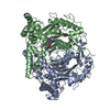
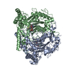
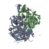
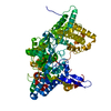
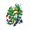
| Title | Crystal structure of L-methionine decarboxylase from Streptomyces sp.590 in complexed with 3-methlythiopropylamine (geminal diamine form). | ||||||
 Components Components | L-methionine decarboxylase | ||||||
 Keywords Keywords | LYASE / decarboxylase / PLP-Dependent Enzymes | ||||||
| Function / homology | Pyridoxal phosphate-dependent transferase, major domain / Pyridoxal phosphate-dependent transferase / Chem-G0F / L-methionine decarboxylase Function and homology information Function and homology information | ||||||
| Biological species |  Streptomyces sp. 590 KI-2014 (bacteria) Streptomyces sp. 590 KI-2014 (bacteria) | ||||||
| Method |  X-RAY DIFFRACTION / SYNCHROTRON / MOLECULAR REPLACEMENT / Resolution: 1.8 Å X-RAY DIFFRACTION / SYNCHROTRON / MOLECULAR REPLACEMENT / Resolution: 1.8 Å | ||||||
 Authors Authors | Okawa, A. / Shiba, T. / Hayashi, M. / Onoue, Y. / Murota, M. / Sato, D. / Inagaki, J. / Tamura, T. / Harada, S. / Inagaki, K. | ||||||
 Citation Citation | Journal: Protein Sci. / Year: 2021 Title: Structural basis for substrate specificity of l-methionine decarboxylase. Authors: Okawa, A. / Shiba, T. / Hayashi, M. / Onoue, Y. / Murota, M. / Sato, D. / Inagaki, J. / Tamura, T. / Harada, S. / Inagaki, K. | ||||||
| History |
|
- Structure visualization
| Structure viewer | Molecule: MolmilJmol/JSmol |
|---|
- Downloads & links
-Download
| PDBx/mmCIF format | 7cim.cif.gz | 218 KB | Display | PDBx/mmCIF format |
|---|---|---|---|---|
| PDB format | pdb7cim.ent.gz | 172.7 KB | Display | PDB format |
| PDBx/mmJSON format | 7cim.json.gz | Tree view | PDBx/mmJSON format | |
| Others |  Other downloads Other downloads |
-Validation report
| Summary document | 7cim_validation.pdf.gz | 1.1 MB | Display | wwPDB validaton report |
|---|---|---|---|---|
| Full document | 7cim_full_validation.pdf.gz | 1.1 MB | Display | |
| Data in XML | 7cim_validation.xml.gz | 39.7 KB | Display | |
| Data in CIF | 7cim_validation.cif.gz | 56.3 KB | Display | |
| Arichive directory | https://data.pdbj.org/pub/pdb/validation_reports/ci/7cimftp://data.pdbj.org/pub/pdb/validation_reports/ci/7cim | HTTPS FTP |
-Related structure data
| Related structure data |  7cifSC  7cigC  7ciiC  7cijC S: Starting model for refinement C: citing same article ( |
|---|---|
| Similar structure data |


-Links
 PDBj
PDBj- Assembly
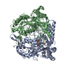
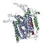
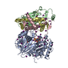
| Deposited unit | 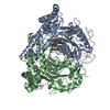
| ||||||||
|---|---|---|---|---|---|---|---|---|---|
| 1 |
| ||||||||
| Unit cell |
|
-Components
| #1: Protein | Mass: 61477.098 Da / Num. of mol.: 2 Source method: isolated from a genetically manipulated source Source: (gene. exp.) Streptomyces sp. 590 KI-2014 (bacteria)Plasmid: pET-52b / Production host: #2: Chemical | 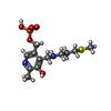  Mass: 336.344 Da / Num. of mol.: 2 / Source method: obtained synthetically / Formula: C12H21N2O5PS / Feature type: SUBJECT OF INVESTIGATION Mass: 336.344 Da / Num. of mol.: 2 / Source method: obtained synthetically / Formula: C12H21N2O5PS / Feature type: SUBJECT OF INVESTIGATION#3: Water | ChemComp-HOH / |  Mass: 18.015 Da / Num. of mol.: 271 / Source method: isolated from a natural source / Formula: H2O Mass: 18.015 Da / Num. of mol.: 271 / Source method: isolated from a natural source / Formula: H2OHas ligand of interest | Y | |
|---|
-Experimental details
-Experiment
| Experiment | Method: X-RAY DIFFRACTION / Number of used crystals: 1 |
|---|
- Sample preparation
| Crystal | Density Matthews: 2.15 Å3/Da / Density % sol: 42.66 % |
|---|---|
| Crystal grow | Temperature: 293 K / Method: vapor diffusion, hanging drop / pH: 6 / Details: 0.1 M MMT buffer pH 6.0, 23.5 % (w/v) PEG1500 / PH range: 5.6-6.4 |
-Data collection
| Diffraction | Mean temperature: 100 K / Serial crystal experiment: N |
|---|---|
| Diffraction source | Source: SYNCHROTRON / Site: SPring-8  / Beamline: BL44XU / Wavelength: 0.9 Å / Beamline: BL44XU / Wavelength: 0.9 Å |
| Detector | Type: DECTRIS EIGER X 16M / Detector: PIXEL / Date: Jun 27, 2019 |
| Radiation | Monochromator: Si(111) / Protocol: SINGLE WAVELENGTH / Monochromatic (M) / Laue (L): M / Scattering type: x-ray |
| Radiation wavelength | Wavelength: 0.9 Å / Relative weight: 1 |
| Reflection | Resolution: 1.8→50 Å / Num. obs: 88135 / % possible obs: 98.1 % / Redundancy: 2.6 % / CC1/2: 0.996 / Rmerge(I) obs: 0.068 / Rpim(I) all: 0.084 / Net I/σ(I): 8.3 |
| Reflection shell | Resolution: 1.8→1.91 Å / Redundancy: 2.4 % / Rmerge(I) obs: 0.419 / Mean I/σ(I) obs: 2 / Num. unique obs: 27664 / CC1/2: 0.87 / Rpim(I) all: 0.519 / % possible all: 95.9 |
- Processing
| Software |
| ||||||||||||||||||||||||||||||||||||||||||||||||||||||||||||||||||||||||||||||||||||||||||||||||||||||||||||||||||||||||||||||||||||||||||||||||||||||||||||||||||||||||||||||||||||||
|---|---|---|---|---|---|---|---|---|---|---|---|---|---|---|---|---|---|---|---|---|---|---|---|---|---|---|---|---|---|---|---|---|---|---|---|---|---|---|---|---|---|---|---|---|---|---|---|---|---|---|---|---|---|---|---|---|---|---|---|---|---|---|---|---|---|---|---|---|---|---|---|---|---|---|---|---|---|---|---|---|---|---|---|---|---|---|---|---|---|---|---|---|---|---|---|---|---|---|---|---|---|---|---|---|---|---|---|---|---|---|---|---|---|---|---|---|---|---|---|---|---|---|---|---|---|---|---|---|---|---|---|---|---|---|---|---|---|---|---|---|---|---|---|---|---|---|---|---|---|---|---|---|---|---|---|---|---|---|---|---|---|---|---|---|---|---|---|---|---|---|---|---|---|---|---|---|---|---|---|---|---|---|---|
| Refinement | Method to determine structure: MOLECULAR REPLACEMENT Starting model: 7CIF Resolution: 1.8→19.96 Å / Cor.coef. Fo:Fc: 0.959 / Cor.coef. Fo:Fc free: 0.943 / SU B: 4.007 / SU ML: 0.118 / Cross valid method: THROUGHOUT / ESU R: 0.151 / ESU R Free: 0.138 / Stereochemistry target values: MAXIMUM LIKELIHOOD / Details: HYDROGENS HAVE BEEN ADDED IN THE RIDING POSITIONS
| ||||||||||||||||||||||||||||||||||||||||||||||||||||||||||||||||||||||||||||||||||||||||||||||||||||||||||||||||||||||||||||||||||||||||||||||||||||||||||||||||||||||||||||||||||||||
| Solvent computation | Ion probe radii: 0.8 Å / Shrinkage radii: 0.8 Å / VDW probe radii: 1.2 Å / Solvent model: MASK | ||||||||||||||||||||||||||||||||||||||||||||||||||||||||||||||||||||||||||||||||||||||||||||||||||||||||||||||||||||||||||||||||||||||||||||||||||||||||||||||||||||||||||||||||||||||
| Displacement parameters | Biso mean: 35.918 Å2
| ||||||||||||||||||||||||||||||||||||||||||||||||||||||||||||||||||||||||||||||||||||||||||||||||||||||||||||||||||||||||||||||||||||||||||||||||||||||||||||||||||||||||||||||||||||||
| Refinement step | Cycle: 1 / Resolution: 1.8→19.96 Å
| ||||||||||||||||||||||||||||||||||||||||||||||||||||||||||||||||||||||||||||||||||||||||||||||||||||||||||||||||||||||||||||||||||||||||||||||||||||||||||||||||||||||||||||||||||||||
| Refine LS restraints |
|