 Movie
Movie Controller


+ Open data

- Basic information
| Entry | Database: PDB / ID: 7ci5 | ||||||
|---|---|---|---|---|---|---|---|
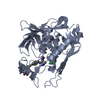
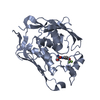
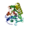
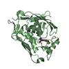
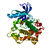
| Title | Crystal structure of P.aeruginosa LpxC in complex with inhibitor | ||||||
 Components Components | UDP-3-O-acyl-N-acetylglucosamine deacetylase | ||||||
 Keywords Keywords | HYDROLASE / UDP-3-O-acyl-N-acetylglucosamine deacetylase / EnvA / LpxC / Pseudomonas aeruginosa | ||||||
| Function / homology |  Function and homology information Function and homology informationUDP-3-O-acyl-N-acetylglucosamine deacetylase / UDP-3-O-acyl-N-acetylglucosamine deacetylase activity / lipid A biosynthetic process / membrane / metal ion binding / cytoplasm Similarity search - Function | ||||||
| Biological species |  Pseudomonas aeruginosa PAO1 (bacteria) Pseudomonas aeruginosa PAO1 (bacteria) | ||||||
| Method |  X-RAY DIFFRACTION / MOLECULAR REPLACEMENT / Resolution: 2.5 Å X-RAY DIFFRACTION / MOLECULAR REPLACEMENT / Resolution: 2.5 Å | ||||||
 Authors Authors | Mima, M. / Baker, L.M. / Surgenor, A. / Robertson, A. | ||||||
 Citation Citation | Journal: J.Med.Chem. / Year: 2020 Title: Fragment-Based Discovery of Novel Non-Hydroxamate LpxC Inhibitors with Antibacterial Activity. Authors: Yamada, Y. / Takashima, H. / Walmsley, D.L. / Ushiyama, F. / Matsuda, Y. / Kanazawa, H. / Yamaguchi-Sasaki, T. / Tanaka-Yamamoto, N. / Yamagishi, J. / Kurimoto-Tsuruta, R. / Ogata, Y. / ...Authors: Yamada, Y. / Takashima, H. / Walmsley, D.L. / Ushiyama, F. / Matsuda, Y. / Kanazawa, H. / Yamaguchi-Sasaki, T. / Tanaka-Yamamoto, N. / Yamagishi, J. / Kurimoto-Tsuruta, R. / Ogata, Y. / Ohtake, N. / Angove, H. / Baker, L. / Harris, R. / Macias, A. / Robertson, A. / Surgenor, A. / Watanabe, H. / Nakano, K. / Mima, M. / Iwamoto, K. / Okada, A. / Takata, I. / Hitaka, K. / Tanaka, A. / Fujita, K. / Sugiyama, H. / Hubbard, R.E. | ||||||
| History |
|
- Structure visualization
| Structure viewer | Molecule: MolmilJmol/JSmol |
|---|
- Downloads & links
-Download
| PDBx/mmCIF format | 7ci5.cif.gz | 238.4 KB | Display | PDBx/mmCIF format |
|---|---|---|---|---|
| PDB format | pdb7ci5.ent.gz | 191.4 KB | Display | PDB format |
| PDBx/mmJSON format | 7ci5.json.gz | Tree view | PDBx/mmJSON format | |
| Others |  Other downloads Other downloads |
-Validation report
| Arichive directory | https://data.pdbj.org/pub/pdb/validation_reports/ci/7ci5ftp://data.pdbj.org/pub/pdb/validation_reports/ci/7ci5 | HTTPS FTP |
|---|
-Related structure data
| Related structure data | 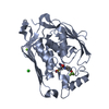 7ci4C 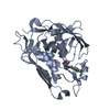 7ci6C  7ci7C 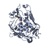 7ci8C 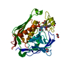 7ci9C  7ciaC  7cibC 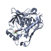 7cicC 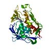 7cidC  7cieC  3uhmS S: Starting model for refinement C: citing same article ( |
|---|---|
| Similar structure data |

-Links
 PDBj
PDBj- Assembly
| Deposited unit | 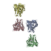
| ||||||||
|---|---|---|---|---|---|---|---|---|---|
| 1 | 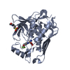
| ||||||||
| 2 | 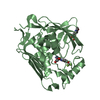
| ||||||||
| 3 | 
| ||||||||
| 4 | 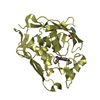
| ||||||||
| Unit cell |
|
-Components
| #1: Protein | Mass: 33146.617 Da / Num. of mol.: 4 / Mutation: C40S Source method: isolated from a genetically manipulated source Source: (gene. exp.) Pseudomonas aeruginosa PAO1 (bacteria) / Strain: PAO1 / Gene: lpxC, envA, PA4406 / Production host: References: UniProt: P47205, UDP-3-O-acyl-N-acetylglucosamine deacetylase #2: Chemical | ChemComp-FXX / (   Mass: 292.211 Da / Num. of mol.: 4 / Source method: obtained synthetically / Formula: C11H11F3N2O4 / Feature type: SUBJECT OF INVESTIGATION Mass: 292.211 Da / Num. of mol.: 4 / Source method: obtained synthetically / Formula: C11H11F3N2O4 / Feature type: SUBJECT OF INVESTIGATION#3: Chemical | ChemComp-MPO /   Mass: 209.263 Da / Num. of mol.: 4 / Source method: obtained synthetically / Formula: C7H15NO4S / Comment: pH buffer*YM Mass: 209.263 Da / Num. of mol.: 4 / Source method: obtained synthetically / Formula: C7H15NO4S / Comment: pH buffer*YM#4: Chemical | ChemComp-ZN /   Mass: 65.409 Da / Num. of mol.: 4 / Source method: obtained synthetically / Formula: Zn Mass: 65.409 Da / Num. of mol.: 4 / Source method: obtained synthetically / Formula: Zn#5: Water | ChemComp-HOH / |  Mass: 18.015 Da / Num. of mol.: 178 / Source method: isolated from a natural source / Formula: H2O Mass: 18.015 Da / Num. of mol.: 178 / Source method: isolated from a natural source / Formula: H2OHas ligand of interest | Y | |
|---|
-Experimental details
-Experiment
| Experiment | Method: X-RAY DIFFRACTION / Number of used crystals: 1 |
|---|
- Sample preparation
| Crystal | Density Matthews: 2.35 Å3/Da / Density % sol: 47.72 % |
|---|---|
| Crystal grow | Temperature: 293 K / Method: vapor diffusion / pH: 7.5 Details: 0.02M 1,6-Hexanediol 0.02M 1-Butanol 0.02M 1,2-Propanediol 0.02M 2-Propanol 0.02M 1,4-Butanediol 0.02M 1,3-Propanediol 0.1M Sodium HEPES - MOPS pH 7.5 20 % v/v PEG 500 MME 10 % w/v PEG 20000 |
-Data collection
| Diffraction | Mean temperature: 100 K / Serial crystal experiment: N |
|---|---|
| Diffraction source | Source: ROTATING ANODE / Type: RIGAKU MICROMAX-007 HF / Wavelength: 1.54184 Å |
| Detector | Type: RIGAKU RAXIS VII / Detector: IMAGE PLATE / Date: May 17, 2016 |
| Radiation | Protocol: SINGLE WAVELENGTH / Monochromatic (M) / Laue (L): M / Scattering type: x-ray |
| Radiation wavelength | Wavelength: 1.54184 Å / Relative weight: 1 |
| Reflection | Resolution: 2.5→22.78 Å / Num. obs: 40242 / % possible obs: 96.4 % / Redundancy: 3.28 % / Rmerge(I) obs: 0.113 / Net I/σ(I): 6.9 |
| Reflection shell | Resolution: 2.5→2.59 Å / Redundancy: 3.21 % / Rmerge(I) obs: 0.347 / Mean I/σ(I) obs: 3 / Num. unique obs: 3920 / % possible all: 94.6 |
- Processing
| Software |
| ||||||||||||||||||||||||||||||||||||||||||||||||||||||||||||||||||||||||||||||||||||||||||||||||||||||||||||||||||||||||||||||||||||||||||||||||||||||||||||||||||||||||||||||||||||||
|---|---|---|---|---|---|---|---|---|---|---|---|---|---|---|---|---|---|---|---|---|---|---|---|---|---|---|---|---|---|---|---|---|---|---|---|---|---|---|---|---|---|---|---|---|---|---|---|---|---|---|---|---|---|---|---|---|---|---|---|---|---|---|---|---|---|---|---|---|---|---|---|---|---|---|---|---|---|---|---|---|---|---|---|---|---|---|---|---|---|---|---|---|---|---|---|---|---|---|---|---|---|---|---|---|---|---|---|---|---|---|---|---|---|---|---|---|---|---|---|---|---|---|---|---|---|---|---|---|---|---|---|---|---|---|---|---|---|---|---|---|---|---|---|---|---|---|---|---|---|---|---|---|---|---|---|---|---|---|---|---|---|---|---|---|---|---|---|---|---|---|---|---|---|---|---|---|---|---|---|---|---|---|---|
| Refinement | Method to determine structure: MOLECULAR REPLACEMENT Starting model: 3UHM Resolution: 2.5→22.78 Å / Cor.coef. Fo:Fc: 0.937 / Cor.coef. Fo:Fc free: 0.875 / SU B: 13.521 / SU ML: 0.291 / Cross valid method: THROUGHOUT / ESU R Free: 0.358 / Stereochemistry target values: MAXIMUM LIKELIHOOD / Details: HYDROGENS HAVE BEEN ADDED IN THE RIDING POSITIONS
| ||||||||||||||||||||||||||||||||||||||||||||||||||||||||||||||||||||||||||||||||||||||||||||||||||||||||||||||||||||||||||||||||||||||||||||||||||||||||||||||||||||||||||||||||||||||
| Solvent computation | Ion probe radii: 0.8 Å / Shrinkage radii: 0.8 Å / VDW probe radii: 1.2 Å / Solvent model: MASK | ||||||||||||||||||||||||||||||||||||||||||||||||||||||||||||||||||||||||||||||||||||||||||||||||||||||||||||||||||||||||||||||||||||||||||||||||||||||||||||||||||||||||||||||||||||||
| Displacement parameters | Biso mean: 38.279 Å2
| ||||||||||||||||||||||||||||||||||||||||||||||||||||||||||||||||||||||||||||||||||||||||||||||||||||||||||||||||||||||||||||||||||||||||||||||||||||||||||||||||||||||||||||||||||||||
| Refinement step | Cycle: 1 / Resolution: 2.5→22.78 Å
| ||||||||||||||||||||||||||||||||||||||||||||||||||||||||||||||||||||||||||||||||||||||||||||||||||||||||||||||||||||||||||||||||||||||||||||||||||||||||||||||||||||||||||||||||||||||
| Refine LS restraints |
|