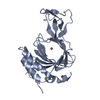
 Movie
Movie Controller


+ Open data

- Basic information
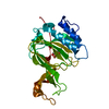
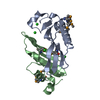
| Entry | Database: PDB / ID: 7chi | ||||||
|---|---|---|---|---|---|---|---|
| Title | crystal structure of pco5 | ||||||
 Components Components | Plant cysteine oxidase 5 | ||||||
 Keywords Keywords | METAL BINDING PROTEIN / oxidase / metal binding / enzyme | ||||||
| Function / homology |  Function and homology information Function and homology informationpeptidyl-cysteine oxidation / detection of hypoxia / cysteine dioxygenase / cysteine dioxygenase activity / cellular response to hypoxia / iron ion binding / nucleus / cytoplasm Similarity search - Function | ||||||
| Biological species |  | ||||||
| Method |  X-RAY DIFFRACTION / SYNCHROTRON / SAD / Resolution: 2.502 Å X-RAY DIFFRACTION / SYNCHROTRON / SAD / Resolution: 2.502 Å | ||||||
 Authors Authors | Guo, Q. / Xu, C. / Liao, S. | ||||||
 Citation Citation | Journal: J.Struct.Biol. / Year: 2021 Title: Molecular basis for cysteine oxidation by plant cysteine oxidases from Arabidopsis thaliana. Authors: Chen, Z. / Guo, Q. / Wu, G. / Wen, J. / Liao, S. / Xu, C. | ||||||
| History |
|
- Structure visualization
| Structure viewer | Molecule: MolmilJmol/JSmol |
|---|
- Downloads & links
-Download
| PDBx/mmCIF format | 7chi.cif.gz | 102.8 KB | Display | PDBx/mmCIF format |
|---|---|---|---|---|
| PDB format | pdb7chi.ent.gz | 77.5 KB | Display | PDB format |
| PDBx/mmJSON format | 7chi.json.gz | Tree view | PDBx/mmJSON format | |
| Others |  Other downloads Other downloads |
-Validation report
| Summary document | 7chi_validation.pdf.gz | 891.2 KB | Display | wwPDB validaton report |
|---|---|---|---|---|
| Full document | 7chi_full_validation.pdf.gz | 892.3 KB | Display | |
| Data in XML | 7chi_validation.xml.gz | 10.7 KB | Display | |
| Data in CIF | 7chi_validation.cif.gz | 14 KB | Display | |
| Arichive directory | https://data.pdbj.org/pub/pdb/validation_reports/ch/7chiftp://data.pdbj.org/pub/pdb/validation_reports/ch/7chi | HTTPS FTP |
-Related structure data


-Links
 PDBj
PDBj- Assembly
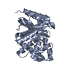
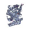
| Deposited unit | 
| ||||||||
|---|---|---|---|---|---|---|---|---|---|
| 1 |
| ||||||||
| Unit cell |
| ||||||||
| Components on special symmetry positions |
|
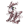
-Components
| #1: Protein | Mass: 27778.576 Da / Num. of mol.: 1 Source method: isolated from a genetically manipulated source Details: SF file contains Friedel pairs. / Source: (gene. exp.) Production host:  References: UniProt: Q9LXT4, cysteine dioxygenase |
|---|---|
| #2: Chemical | ChemComp-FE /   Mass: 55.845 Da / Num. of mol.: 1 / Source method: obtained synthetically / Formula: Fe / Feature type: SUBJECT OF INVESTIGATION Mass: 55.845 Da / Num. of mol.: 1 / Source method: obtained synthetically / Formula: Fe / Feature type: SUBJECT OF INVESTIGATION |
| #3: Water | ChemComp-HOH /  Mass: 18.015 Da / Num. of mol.: 62 / Source method: isolated from a natural source / Formula: H2O Mass: 18.015 Da / Num. of mol.: 62 / Source method: isolated from a natural source / Formula: H2O |
| Has ligand of interest | Y |
| Has protein modification | Y |
-Experimental details
-Experiment
| Experiment | Method: X-RAY DIFFRACTION / Number of used crystals: 1 |
|---|
- Sample preparation
| Crystal | Density Matthews: 2.65 Å3/Da / Density % sol: 53.63 % |
|---|---|
| Crystal grow | Temperature: 291.1 K / Method: vapor diffusion, sitting drop / Details: 12% PEG20000, 0.1M MES monohydrate pH 6.5 |
-Data collection
| Diffraction | Mean temperature: 100 K / Serial crystal experiment: N |
|---|---|
| Diffraction source | Source: SYNCHROTRON / Site: SSRF  / Beamline: BL18U1 / Wavelength: 0.9792 Å / Beamline: BL18U1 / Wavelength: 0.9792 Å |
| Detector | Type: DECTRIS PILATUS3 6M / Detector: PIXEL / Date: Jun 19, 2020 |
| Radiation | Protocol: SINGLE WAVELENGTH / Monochromatic (M) / Laue (L): M / Scattering type: x-ray |
| Radiation wavelength | Wavelength: 0.9792 Å / Relative weight: 1 |
| Reflection | Resolution: 2.5→50 Å / Num. obs: 19655 / % possible obs: 100 % / Redundancy: 24.4 % / CC1/2: 0.985 / Rmerge(I) obs: 0.135 / Net I/σ(I): 32.6 |
| Reflection shell | Resolution: 2.5→2.59 Å / Rmerge(I) obs: 1.625 / Mean I/σ(I) obs: 4 / Num. unique obs: 1064 / CC1/2: 0.894 |
- Processing
| Software |
| ||||||||||||||||||||||||||||||||||||||||||||||||||||||||||||||||||||||||||||||||||||||||||||||||||||
|---|---|---|---|---|---|---|---|---|---|---|---|---|---|---|---|---|---|---|---|---|---|---|---|---|---|---|---|---|---|---|---|---|---|---|---|---|---|---|---|---|---|---|---|---|---|---|---|---|---|---|---|---|---|---|---|---|---|---|---|---|---|---|---|---|---|---|---|---|---|---|---|---|---|---|---|---|---|---|---|---|---|---|---|---|---|---|---|---|---|---|---|---|---|---|---|---|---|---|---|---|---|
| Refinement | Method to determine structure: SAD / Resolution: 2.502→24.717 Å / SU ML: 0.28 / Cross valid method: THROUGHOUT / σ(F): 1.35 / Phase error: 22.31 / Stereochemistry target values: ML
| ||||||||||||||||||||||||||||||||||||||||||||||||||||||||||||||||||||||||||||||||||||||||||||||||||||
| Solvent computation | Shrinkage radii: 0.9 Å / VDW probe radii: 1.11 Å / Solvent model: FLAT BULK SOLVENT MODEL | ||||||||||||||||||||||||||||||||||||||||||||||||||||||||||||||||||||||||||||||||||||||||||||||||||||
| Displacement parameters | Biso max: 109.57 Å2 / Biso mean: 43.3026 Å2 / Biso min: 14.18 Å2 | ||||||||||||||||||||||||||||||||||||||||||||||||||||||||||||||||||||||||||||||||||||||||||||||||||||
| Refinement step | Cycle: final / Resolution: 2.502→24.717 Å
| ||||||||||||||||||||||||||||||||||||||||||||||||||||||||||||||||||||||||||||||||||||||||||||||||||||
| LS refinement shell | Refine-ID: X-RAY DIFFRACTION / Rfactor Rfree error: 0 / % reflection obs: 100 %
| ||||||||||||||||||||||||||||||||||||||||||||||||||||||||||||||||||||||||||||||||||||||||||||||||||||
| Refinement TLS params. | Method: refined / Refine-ID: X-RAY DIFFRACTION
| ||||||||||||||||||||||||||||||||||||||||||||||||||||||||||||||||||||||||||||||||||||||||||||||||||||
| Refinement TLS group |
|
