 Movie
Movie Controller


+ Open data

- Basic information
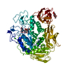
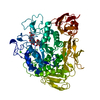
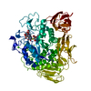
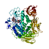
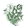
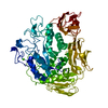
| Entry | Database: PDB / ID: 7cgt | ||||||
|---|---|---|---|---|---|---|---|
| Title | RAMEB COMPLEX OF CYCLODEXTRIN GLYCOSYLTRANSFERASE MUTANT | ||||||
 Components Components | CYCLODEXTRIN GLYCOSYLTRANSFERASE | ||||||
 Keywords Keywords | GLYCOSYLTRANSFERASE / STARCH DEGRADATION / CYCLODEXTRIN | ||||||
| Function / homology |  Function and homology information Function and homology informationcyclomaltodextrin glucanotransferase / cyclomaltodextrin glucanotransferase activity / starch binding / alpha-amylase activity / carbohydrate metabolic process / extracellular region / metal ion binding Similarity search - Function | ||||||
| Biological species |  Bacillus circulans (bacteria) Bacillus circulans (bacteria) | ||||||
| Method |  X-RAY DIFFRACTION / X-PLOR WITH WILD-TYPE MODEL / Resolution: 3 Å X-RAY DIFFRACTION / X-PLOR WITH WILD-TYPE MODEL / Resolution: 3 Å | ||||||
 Authors Authors | Parsiegla, G. / Schulz, G.E. | ||||||
 Citation Citation | Journal: Eur.J.Biochem. / Year: 1998 Title: Substrate binding to a cyclodextrin glycosyltransferase and mutations increasing the gamma-cyclodextrin production. Authors: Parsiegla, G. / Schmidt, A.K. / Schulz, G.E. #1: Journal: Biochemistry / Year: 1998Title: Structure of Cyclodextrin Glycosyltransferase Complexed with a Derivative of its Main Product Beta-Cyclodextrin Authors: Schmidt, A.K. / Cottaz, S. / Driguez, H. / Schulz, G.E. #2: Journal: Biochemistry / Year: 1992Title: Catalytic Center of Cyclodextrin Glycosyltransferase Derived from X-Ray Structure Analysis Combined with Site-Directed Mutagenesis Authors: Klein, C. / Hollender, J. / Bender, H. / Schulz, G.E. #3: Journal: J.Mol.Biol. / Year: 1991Title: Structure of Cyclodextrin Glycosyltransferase Refined at 2.0 A Resolution Authors: Klein, C. / Schulz, G.E. #4: Journal: Appl.Microbiol.Biotechnol. / Year: 1990Title: Molecular Cloning, Nucleotide Sequence and Expression in Escherichia Coli of the Beta-Cyclodextrin Glycosyltransferase Gene from Bacillus Circulans Strain No. 8 Authors: Nitschke, L. / Heeger, K. / Bender, H. / Schulz, G.E. | ||||||
| History |
|
- Structure visualization
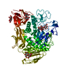
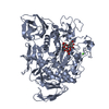
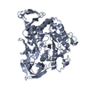
| Structure viewer | Molecule: MolmilJmol/JSmol |
|---|
- Downloads & links
-Download
| PDBx/mmCIF format | 7cgt.cif.gz | 132.4 KB | Display | PDBx/mmCIF format |
|---|---|---|---|---|
| PDB format | pdb7cgt.ent.gz | 103.7 KB | Display | PDB format |
| PDBx/mmJSON format | 7cgt.json.gz | Tree view | PDBx/mmJSON format | |
| Others |  Other downloads Other downloads |
-Validation report
| Summary document | 7cgt_validation.pdf.gz | 417.4 KB | Display | wwPDB validaton report |
|---|---|---|---|---|
| Full document | 7cgt_full_validation.pdf.gz | 433.8 KB | Display | |
| Data in XML | 7cgt_validation.xml.gz | 26.6 KB | Display | |
| Data in CIF | 7cgt_validation.cif.gz | 37 KB | Display | |
| Arichive directory | https://data.pdbj.org/pub/pdb/validation_reports/cg/7cgtftp://data.pdbj.org/pub/pdb/validation_reports/cg/7cgt | HTTPS FTP |
-Related structure data


-Links
 PDBj
PDBj


- Assembly
| Deposited unit | 
| ||||||||
|---|---|---|---|---|---|---|---|---|---|
| 1 |
| ||||||||
| Unit cell |
|
-Components
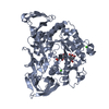
| #1: Protein | Mass: 74521.031 Da / Num. of mol.: 1 / Mutation: D229A Source method: isolated from a genetically manipulated source Source: (gene. exp.) Bacillus circulans (bacteria) / Strain: 8 / Cellular location: EXTRACELLULAR / Production host: References: UniProt: P30920, cyclomaltodextrin glucanotransferase | ||||
|---|---|---|---|---|---|
| #2: Chemical |   Mass: 40.078 Da / Num. of mol.: 2 / Source method: obtained synthetically / Formula: Ca Mass: 40.078 Da / Num. of mol.: 2 / Source method: obtained synthetically / Formula: Ca#3: Water | ChemComp-HOH / |  Mass: 18.015 Da / Num. of mol.: 99 / Source method: isolated from a natural source / Formula: H2O Mass: 18.015 Da / Num. of mol.: 99 / Source method: isolated from a natural source / Formula: H2OHas protein modification | Y | |
-Experimental details
-Experiment
| Experiment | Method: X-RAY DIFFRACTION |
|---|
- Sample preparation
| Crystal | Density Matthews: 3.79 Å3/Da / Density % sol: 67.57 % | |||||||||||||||||||||||||||||||||||||||||||||
|---|---|---|---|---|---|---|---|---|---|---|---|---|---|---|---|---|---|---|---|---|---|---|---|---|---|---|---|---|---|---|---|---|---|---|---|---|---|---|---|---|---|---|---|---|---|---|
| Crystal grow | pH: 6.9 Details: pH 6.9 THE SUBMITTED PDB FILE RESULTED FROM A SOAK OF CGTASE MUTANT D229A WITH A 1.8 TIMES RANDOMLY METHYLATED BETA-CYCLODEXTRIN (RAMEB). THE STRUCTURE WAS REFINED WITH A NON-METHYLATED BETA- ...Details: pH 6.9 THE SUBMITTED PDB FILE RESULTED FROM A SOAK OF CGTASE MUTANT D229A WITH A 1.8 TIMES RANDOMLY METHYLATED BETA-CYCLODEXTRIN (RAMEB). THE STRUCTURE WAS REFINED WITH A NON-METHYLATED BETA-CYCLODEXTRIN MODEL WHICH IS NOT INCLUDED IN THE PDB FILE BECAUSE OF INSUFFICIENT DENSITY. | |||||||||||||||||||||||||||||||||||||||||||||
| Crystal grow | *PLUS pH: 6.7 / Method: vapor diffusion, hanging drop / Details: Hofmann, B.E., (1989) J.Mol.Biol., 209, 793. | |||||||||||||||||||||||||||||||||||||||||||||
| Components of the solutions | *PLUS
|
-Data collection
| Diffraction | Mean temperature: 298 K |
|---|---|
| Diffraction source | Source: ROTATING ANODE / Type: RIGAKU RUH2R / Wavelength: 1.5418 |
| Detector | Type: SIEMENS / Detector: AREA DETECTOR / Date: Aug 1, 1993 |
| Radiation | Monochromatic (M) / Laue (L): M / Scattering type: x-ray |
| Radiation wavelength | Wavelength: 1.5418 Å / Relative weight: 1 |
| Reflection | Resolution: 3→35.6 Å / Num. obs: 18086 / % possible obs: 82.5 % |
| Reflection shell | Highest resolution: 3 Å / % possible all: 58 |
- Processing
| Software |
| ||||||||||||||||||||||||||||||||||||||||||||||||||||||||||||
|---|---|---|---|---|---|---|---|---|---|---|---|---|---|---|---|---|---|---|---|---|---|---|---|---|---|---|---|---|---|---|---|---|---|---|---|---|---|---|---|---|---|---|---|---|---|---|---|---|---|---|---|---|---|---|---|---|---|---|---|---|---|
| Refinement | Method to determine structure: X-PLOR WITH WILD-TYPE MODEL / Resolution: 3→10 Å / Cross valid method: THROUGHOUT Details: THE STRUCTURE WAS REFINED WITH A BETA-CYCLODEXTRIN MODEL WHICH IS NOT INCLUDED IN THE SUBMITTED PDB FILE.
| ||||||||||||||||||||||||||||||||||||||||||||||||||||||||||||
| Displacement parameters | Biso mean: 13.1 Å2 | ||||||||||||||||||||||||||||||||||||||||||||||||||||||||||||
| Refinement step | Cycle: LAST / Resolution: 3→10 Å
| ||||||||||||||||||||||||||||||||||||||||||||||||||||||||||||
| Refine LS restraints |
| ||||||||||||||||||||||||||||||||||||||||||||||||||||||||||||
| Xplor file |
|
