 Movie
Movie Controller

[English] 日本語
 Yorodumi
- PDB-7cg8: Structure of the sensor domain (short construct) of the anti-sigm... -
+ Open data

- Basic information
| Entry | Database: PDB / ID: 7cg8 | ||||||
|---|---|---|---|---|---|---|---|
| Title | Structure of the sensor domain (short construct) of the anti-sigma factor RsgI4 in Pseudobacteroides cellulosolvens | ||||||
 Components Components | Anti-sigma factor RsgI, N-terminal | ||||||
 Keywords Keywords | TRANSCRIPTION / Sugar binding protein | ||||||
| Function / homology | Anti-sigma factor RsgI4, sensor domain / : / Anti-sigma factor RsgI-like central domain / Anti-sigma factor RsgI, N-terminal / Anti-sigma factor N-terminus / RsgI N-terminal anti-sigma domain profile. / plasma membrane / ACETATE ION / Anti-sigma factor RsgI, N-terminal Function and homology information Function and homology information | ||||||
| Biological species |  Pseudobacteroides cellulosolvens ATCC 35603 = DSM 2933 (bacteria) Pseudobacteroides cellulosolvens ATCC 35603 = DSM 2933 (bacteria) | ||||||
| Method |  X-RAY DIFFRACTION / SYNCHROTRON / MOLECULAR REPLACEMENT / Resolution: 1.5 Å X-RAY DIFFRACTION / SYNCHROTRON / MOLECULAR REPLACEMENT / Resolution: 1.5 Å | ||||||
 Authors Authors | Dong, S. / Feng, Y. | ||||||
| Funding support |  China, 1items China, 1items
| ||||||
 Citation Citation | Journal: Protein Sci. / Year: 2024 Title: Unique Fn3-like biosensor in sigma I /anti-sigma I factors for regulatory expression of major cellulosomal scaffoldins in Pseudobacteroides cellulosolvens. Authors: Dong, S. / Chen, C. / Li, J. / Liu, Y.J. / Bayer, E.A. / Lamed, R. / Mizrahi, I. / Cui, Q. / Feng, Y. | ||||||
| History |
|
- Structure visualization
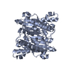
| Structure viewer | Molecule: MolmilJmol/JSmol |
|---|
- Downloads & links
-Download
| PDBx/mmCIF format | 7cg8.cif.gz | 222.8 KB | Display | PDBx/mmCIF format |
|---|---|---|---|---|
| PDB format | pdb7cg8.ent.gz | 145.4 KB | Display | PDB format |
| PDBx/mmJSON format | 7cg8.json.gz | Tree view | PDBx/mmJSON format | |
| Others |  Other downloads Other downloads |
-Validation report
| Arichive directory | https://data.pdbj.org/pub/pdb/validation_reports/cg/7cg8ftp://data.pdbj.org/pub/pdb/validation_reports/cg/7cg8 | HTTPS FTP |
|---|
-Related structure data
| Related structure data | 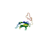 7cg1C  7cg5SC S: Starting model for refinement C: citing same article ( |
|---|---|
| Similar structure data |


-Links
 PDBj
PDBj- Assembly
| Deposited unit | 
| ||||||||||||
|---|---|---|---|---|---|---|---|---|---|---|---|---|---|
| 1 |
| ||||||||||||
| Unit cell |
|
-Components
| #1: Protein | Mass: 11445.965 Da / Num. of mol.: 4 / Fragment: sensor domain Source method: isolated from a genetically manipulated source Source: (gene. exp.) Pseudobacteroides cellulosolvens ATCC 35603 = DSM 2933 (bacteria)Gene: Bccel_2225 / Plasmid: pET28a-SMT3 / Production host: #2: Chemical |   Mass: 59.044 Da / Num. of mol.: 3 / Source method: obtained synthetically / Formula: C2H3O2 / Feature type: SUBJECT OF INVESTIGATION Mass: 59.044 Da / Num. of mol.: 3 / Source method: obtained synthetically / Formula: C2H3O2 / Feature type: SUBJECT OF INVESTIGATION#3: Chemical |   Mass: 1221.461 Da / Num. of mol.: 2 / Source method: obtained synthetically / Formula: C55H112O28 / Feature type: SUBJECT OF INVESTIGATION / Comment: precipitant*YM Mass: 1221.461 Da / Num. of mol.: 2 / Source method: obtained synthetically / Formula: C55H112O28 / Feature type: SUBJECT OF INVESTIGATION / Comment: precipitant*YM#4: Water | ChemComp-HOH / |  Mass: 18.015 Da / Num. of mol.: 580 / Source method: isolated from a natural source / Formula: H2O Mass: 18.015 Da / Num. of mol.: 580 / Source method: isolated from a natural source / Formula: H2OHas ligand of interest | Y | Has protein modification | N | |
|---|
-Experimental details
-Experiment
| Experiment | Method: X-RAY DIFFRACTION / Number of used crystals: 1 |
|---|
- Sample preparation
| Crystal | Density Matthews: 2.13 Å3/Da / Density % sol: 42.18 % |
|---|---|
| Crystal grow | Temperature: 291 K / Method: vapor diffusion, hanging drop / pH: 6.5 Details: 0.2 M ammonium acetate, 0.1 M Bis-Tris (pH 6.5), 25% PEG3350 |
-Data collection
| Diffraction | Mean temperature: 100 K / Serial crystal experiment: N |
|---|---|
| Diffraction source | Source: SYNCHROTRON / Site: SSRF / Beamline: BL17U1 / Wavelength: 0.979183 Å |
| Detector | Type: DECTRIS EIGER X 16M / Detector: PIXEL / Date: Oct 27, 2019 |
| Radiation | Protocol: SINGLE WAVELENGTH / Monochromatic (M) / Laue (L): M / Scattering type: x-ray |
| Radiation wavelength | Wavelength: 0.979183 Å / Relative weight: 1 |
| Reflection | Resolution: 1.5→47.96 Å / Num. obs: 120016 / % possible obs: 99.3 % / Redundancy: 3.48 % / Biso Wilson estimate: 15.37 Å2 / CC1/2: 0.998 / Rrim(I) all: 0.074 / Net I/σ(I): 11.44 |
| Reflection shell | Resolution: 1.5→1.54 Å / Mean I/σ(I) obs: 1.8 / Num. unique obs: 8817 / CC1/2: 0.664 / Rrim(I) all: 0.837 / % possible all: 98.6 |
- Processing
| Software |
| ||||||||||||||||||||||||||||||||||||||||||||||||||||||||||||||||||||||||||||||||||||||||||||||||||||||||||||||||||||||||||||||||||||||||||||||||||||||||||||||||||||||||||||||||||||||||||||||||||||
|---|---|---|---|---|---|---|---|---|---|---|---|---|---|---|---|---|---|---|---|---|---|---|---|---|---|---|---|---|---|---|---|---|---|---|---|---|---|---|---|---|---|---|---|---|---|---|---|---|---|---|---|---|---|---|---|---|---|---|---|---|---|---|---|---|---|---|---|---|---|---|---|---|---|---|---|---|---|---|---|---|---|---|---|---|---|---|---|---|---|---|---|---|---|---|---|---|---|---|---|---|---|---|---|---|---|---|---|---|---|---|---|---|---|---|---|---|---|---|---|---|---|---|---|---|---|---|---|---|---|---|---|---|---|---|---|---|---|---|---|---|---|---|---|---|---|---|---|---|---|---|---|---|---|---|---|---|---|---|---|---|---|---|---|---|---|---|---|---|---|---|---|---|---|---|---|---|---|---|---|---|---|---|---|---|---|---|---|---|---|---|---|---|---|---|---|---|---|
| Refinement | Method to determine structure: MOLECULAR REPLACEMENT Starting model: 7CG5 Resolution: 1.5→33.49 Å / SU ML: 0.1558 / Cross valid method: FREE R-VALUE / σ(F): 1.35 / Phase error: 20.121 Stereochemistry target values: GeoStd + Monomer Library + CDL v1.2
| ||||||||||||||||||||||||||||||||||||||||||||||||||||||||||||||||||||||||||||||||||||||||||||||||||||||||||||||||||||||||||||||||||||||||||||||||||||||||||||||||||||||||||||||||||||||||||||||||||||
| Solvent computation | Shrinkage radii: 0.9 Å / VDW probe radii: 1.11 Å / Solvent model: FLAT BULK SOLVENT MODEL | ||||||||||||||||||||||||||||||||||||||||||||||||||||||||||||||||||||||||||||||||||||||||||||||||||||||||||||||||||||||||||||||||||||||||||||||||||||||||||||||||||||||||||||||||||||||||||||||||||||
| Displacement parameters | Biso mean: 22.13 Å2 | ||||||||||||||||||||||||||||||||||||||||||||||||||||||||||||||||||||||||||||||||||||||||||||||||||||||||||||||||||||||||||||||||||||||||||||||||||||||||||||||||||||||||||||||||||||||||||||||||||||
| Refinement step | Cycle: LAST / Resolution: 1.5→33.49 Å
| ||||||||||||||||||||||||||||||||||||||||||||||||||||||||||||||||||||||||||||||||||||||||||||||||||||||||||||||||||||||||||||||||||||||||||||||||||||||||||||||||||||||||||||||||||||||||||||||||||||
| Refine LS restraints |
| ||||||||||||||||||||||||||||||||||||||||||||||||||||||||||||||||||||||||||||||||||||||||||||||||||||||||||||||||||||||||||||||||||||||||||||||||||||||||||||||||||||||||||||||||||||||||||||||||||||
| LS refinement shell |
|
