 Movie
Movie Controller


+ Open data

- Basic information
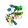
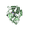
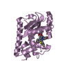
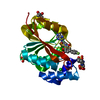
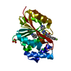
| Entry | Database: PDB / ID: 7cfe | ||||||
|---|---|---|---|---|---|---|---|
| Title | Crystal structure of RsmG methyltransferase of M. tuberculosis | ||||||
 Components Components | Ribosomal RNA small subunit methyltransferase G | ||||||
 Keywords Keywords | TRANSFERASE / Rossmann fold / RsmG / Rv3919c / M. tuberculosis / gidB | ||||||
| Function / homology |  Function and homology information Function and homology informationrRNA (guanine-N7-)-methyltransferase activity / Transferases; Transferring one-carbon groups; Methyltransferases / plasma membrane / cytosol Similarity search - Function | ||||||
| Biological species |   Mycobacterium tuberculosis (bacteria) Mycobacterium tuberculosis (bacteria) | ||||||
| Method |  X-RAY DIFFRACTION / SAD / Resolution: 2.02 Å X-RAY DIFFRACTION / SAD / Resolution: 2.02 Å | ||||||
 Authors Authors | Bijpuria, S. / Maurya, A. / Kumar, P. / Sharma, R. / Taneja, B. | ||||||
| Funding support |  India, 1items India, 1items
| ||||||
 Citation Citation | Journal: To Be Published Title: Crystal structure of RsmG methyltransferase of M. tuberculosis Authors: Bijpuria, S. / Maurya, A. / Kumar, P. / Sharma, R. / Taneja, B. | ||||||
| History |
|
- Structure visualization
| Structure viewer | Molecule: MolmilJmol/JSmol |
|---|
- Downloads & links
-Download
| PDBx/mmCIF format | 7cfe.cif.gz | 60.8 KB | Display | PDBx/mmCIF format |
|---|---|---|---|---|
| PDB format | pdb7cfe.ent.gz | 41.4 KB | Display | PDB format |
| PDBx/mmJSON format | 7cfe.json.gz | Tree view | PDBx/mmJSON format | |
| Others |  Other downloads Other downloads |
-Validation report
| Summary document | 7cfe_validation.pdf.gz | 731.1 KB | Display | wwPDB validaton report |
|---|---|---|---|---|
| Full document | 7cfe_full_validation.pdf.gz | 733.1 KB | Display | |
| Data in XML | 7cfe_validation.xml.gz | 12.1 KB | Display | |
| Data in CIF | 7cfe_validation.cif.gz | 17 KB | Display | |
| Arichive directory | https://data.pdbj.org/pub/pdb/validation_reports/cf/7cfeftp://data.pdbj.org/pub/pdb/validation_reports/cf/7cfe | HTTPS FTP |
-Related structure data
| Similar structure data |
|---|


-Links
 PDBj
PDBj

- Assembly
| Deposited unit | 
| ||||||||
|---|---|---|---|---|---|---|---|---|---|
| 1 |
| ||||||||
| Unit cell |
|
-Components
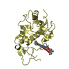
| #1: Protein | Mass: 24895.830 Da / Num. of mol.: 1 Source method: isolated from a genetically manipulated source Source: (gene. exp.) Mycobacterium tuberculosis (strain ATCC 25618 / H37Rv) (bacteria)Strain: ATCC 25618 / H37Rv / Gene: rsmG, gidB, Rv3919c, MTV028.10c / Production host: Mycolicibacterium smegmatis (bacteria)References: UniProt: P9WGW9, Transferases; Transferring one-carbon groups; Methyltransferases |
|---|---|
| #2: Chemical | ChemComp-SFG /   Mass: 381.387 Da / Num. of mol.: 1 / Source method: obtained synthetically / Formula: C15H23N7O5 / Feature type: SUBJECT OF INVESTIGATION Mass: 381.387 Da / Num. of mol.: 1 / Source method: obtained synthetically / Formula: C15H23N7O5 / Feature type: SUBJECT OF INVESTIGATION |
| #3: Chemical | ChemComp-PO4 /   Mass: 94.971 Da / Num. of mol.: 1 / Source method: obtained synthetically / Formula: PO4 Mass: 94.971 Da / Num. of mol.: 1 / Source method: obtained synthetically / Formula: PO4 |
| #4: Chemical | ChemComp-IPA /   Mass: 60.095 Da / Num. of mol.: 1 / Source method: obtained synthetically / Formula: C3H8O Mass: 60.095 Da / Num. of mol.: 1 / Source method: obtained synthetically / Formula: C3H8O |
| #5: Water | ChemComp-HOH /  Mass: 18.015 Da / Num. of mol.: 154 / Source method: isolated from a natural source / Formula: H2O Mass: 18.015 Da / Num. of mol.: 154 / Source method: isolated from a natural source / Formula: H2O |
| Has ligand of interest | Y |
-Experimental details
-Experiment
| Experiment | Method: X-RAY DIFFRACTION / Number of used crystals: 1 |
|---|
- Sample preparation
| Crystal | Density Matthews: 2.06 Å3/Da / Density % sol: 41.11 % |
|---|---|
| Crystal grow | Temperature: 296 K / Method: vapor diffusion Details: 0.1 M Bis-tris pH 7.5, 0.18 M Na/K phosphate and 28% PEG 3350 |
-Data collection
| Diffraction | Mean temperature: 100 K / Serial crystal experiment: N |
|---|---|
| Diffraction source | Source: ROTATING ANODE / Site: RRCAT INDUS-2 / Beamline: PX-BL21 / Wavelength: 0.97949 Å |
| Detector | Type: MARMOSAIC 225 mm CCD / Detector: CCD / Date: Mar 19, 2019 |
| Radiation | Protocol: SINGLE WAVELENGTH / Monochromatic (M) / Laue (L): M / Scattering type: x-ray |
| Radiation wavelength | Wavelength: 0.97949 Å / Relative weight: 1 |
| Reflection | Resolution: 2.02→27.26 Å / Num. obs: 13090 / % possible obs: 98.7 % / Redundancy: 5.6 % / CC1/2: 0.991 / Net I/σ(I): 6.6 |
| Reflection shell | Resolution: 2.02→2.07 Å / Num. unique obs: 799 / CC1/2: 0.532 |
- Processing
| Software |
| |||||||||||||||||||||||||||||||||||
|---|---|---|---|---|---|---|---|---|---|---|---|---|---|---|---|---|---|---|---|---|---|---|---|---|---|---|---|---|---|---|---|---|---|---|---|---|
| Refinement | Method to determine structure: SAD / Resolution: 2.02→27.26 Å / SU ML: 0.24 / Cross valid method: FREE R-VALUE / σ(F): 1.34 / Phase error: 23.21 / Stereochemistry target values: ML
| |||||||||||||||||||||||||||||||||||
| Solvent computation | Shrinkage radii: 0.9 Å / VDW probe radii: 1.11 Å / Solvent model: FLAT BULK SOLVENT MODEL | |||||||||||||||||||||||||||||||||||
| Refinement step | Cycle: LAST / Resolution: 2.02→27.26 Å
| |||||||||||||||||||||||||||||||||||
| Refine LS restraints |
| |||||||||||||||||||||||||||||||||||
| LS refinement shell |
|
