 Movie
Movie Controller

[English] 日本語
 Yorodumi
- PDB-7cdm: Lysozyme room-temperature structure determined by SS-ROX combined... -
+ Open data

- Basic information
| Entry | Database: PDB / ID: 7cdm | |||||||||
|---|---|---|---|---|---|---|---|---|---|---|
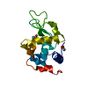
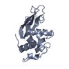
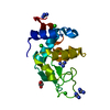
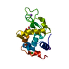
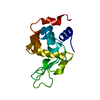
| Title | Lysozyme room-temperature structure determined by SS-ROX combined with HAG method, 42 kGy (4500 images from 2nd half of data set) | |||||||||
 Components Components | Lysozyme C | |||||||||
 Keywords Keywords | HYDROLASE / Chicken egg white / antimicrobial enzyme / N-acetylmuramide glycanhydrolase / lysozyme-like fold | |||||||||
| Function / homology |  Function and homology information Function and homology informationLactose synthesis / Antimicrobial peptides / Neutrophil degranulation / beta-N-acetylglucosaminidase activity / cell wall macromolecule catabolic process / lysozyme / lysozyme activity / killing of cells of another organism / defense response to Gram-negative bacterium / defense response to bacterium ...Lactose synthesis / Antimicrobial peptides / Neutrophil degranulation / beta-N-acetylglucosaminidase activity / cell wall macromolecule catabolic process / lysozyme / lysozyme activity / killing of cells of another organism / defense response to Gram-negative bacterium / defense response to bacterium / defense response to Gram-positive bacterium / endoplasmic reticulum / : / identical protein binding / cytoplasm Similarity search - Function | |||||||||
| Biological species |  | |||||||||
| Method |  X-RAY DIFFRACTION / SYNCHROTRON / FOURIER SYNTHESIS / Resolution: 1.7 Å X-RAY DIFFRACTION / SYNCHROTRON / FOURIER SYNTHESIS / Resolution: 1.7 Å | |||||||||
 Authors Authors | Hasegawa, K. / Baba, S. / Kawamura, T. / Yamamoto, M. / Kumasaka, T. | |||||||||
| Funding support |  Japan, 2items Japan, 2items
| |||||||||
 Citation Citation | Journal: Acta Crystallogr.,Sect.D / Year: 2021 Title: Evaluation of the data-collection strategy for room-temperature micro-crystallography studied by serial synchrotron rotation crystallography combined with the humid air and glue-coating method. Authors: Hasegawa, K. / Baba, S. / Kawamura, T. / Yamamoto, M. / Kumasaka, T. | |||||||||
| History |
|
- Structure visualization
| Structure viewer | Molecule: MolmilJmol/JSmol |
|---|
- Downloads & links
-Download
| PDBx/mmCIF format | 7cdm.cif.gz | 47.7 KB | Display | PDBx/mmCIF format |
|---|---|---|---|---|
| PDB format | pdb7cdm.ent.gz | 26.2 KB | Display | PDB format |
| PDBx/mmJSON format | 7cdm.json.gz | Tree view | PDBx/mmJSON format | |
| Others |  Other downloads Other downloads |
-Validation report
| Arichive directory | https://data.pdbj.org/pub/pdb/validation_reports/cd/7cdmftp://data.pdbj.org/pub/pdb/validation_reports/cd/7cdm | HTTPS FTP |
|---|
-Related structure data
| Related structure data |  7cdkC  7cdnC  7cdoC  7cdpC  7cdqC  7cdrC  7cdsC  7cdtC  7cduC  4etaS S: Starting model for refinement C: citing same article ( |
|---|---|
| Similar structure data |


-Links
 PDBj
PDBj


- Assembly
| Deposited unit | 
| ||||||||||||
|---|---|---|---|---|---|---|---|---|---|---|---|---|---|
| 1 |
| ||||||||||||
| Unit cell |
| ||||||||||||
| Components on special symmetry positions |
|
-Components
| #1: Protein | Mass: 14331.160 Da / Num. of mol.: 1 Source method: isolated from a genetically manipulated source Source: (gene. exp.) |
|---|---|
| #2: Chemical | ChemComp-MLI /   Mass: 102.046 Da / Num. of mol.: 1 / Source method: obtained synthetically / Formula: C3H2O4 Mass: 102.046 Da / Num. of mol.: 1 / Source method: obtained synthetically / Formula: C3H2O4 |
| #3: Chemical | ChemComp-NA /   Mass: 22.990 Da / Num. of mol.: 1 / Source method: obtained synthetically / Formula: Na Mass: 22.990 Da / Num. of mol.: 1 / Source method: obtained synthetically / Formula: Na |
| #4: Water | ChemComp-HOH /  Mass: 18.015 Da / Num. of mol.: 49 / Source method: isolated from a natural source / Formula: H2O Mass: 18.015 Da / Num. of mol.: 49 / Source method: isolated from a natural source / Formula: H2O |
| Has ligand of interest | N |
| Has protein modification | Y |
-Experimental details
-Experiment
| Experiment | Method: X-RAY DIFFRACTION / Number of used crystals: 1 |
|---|
- Sample preparation
| Crystal | Density Matthews: 2.08 Å3/Da / Density % sol: 40.79 % |
|---|---|
| Crystal grow | Temperature: 293 K / Method: batch mode Details: The lysozyme solution (40 mg/ml) in 10 mM Na-acetate, pH 4.6 was mixed with the same volume of 4 M Na-Malonate pH 3.1, 6% w/v PEG6000 and stirred for 20 minutes. The crystal growth was ...Details: The lysozyme solution (40 mg/ml) in 10 mM Na-acetate, pH 4.6 was mixed with the same volume of 4 M Na-Malonate pH 3.1, 6% w/v PEG6000 and stirred for 20 minutes. The crystal growth was stopped by adding 2.4 M Na-Malonate pH 3.1, 3.6 % w/v PEG6000. |
-Data collection
| Diffraction | Mean temperature: 298 K / Serial crystal experiment: Y |
|---|---|
| Diffraction source | Source: SYNCHROTRON / Site: SPring-8 / Beamline: BL41XU / Wavelength: 1 Å |
| Detector | Type: DECTRIS EIGER X 16M / Detector: PIXEL / Date: Nov 13, 2019 |
| Radiation | Protocol: SINGLE WAVELENGTH / Monochromatic (M) / Laue (L): M / Scattering type: x-ray |
| Radiation wavelength | Wavelength: 1 Å / Relative weight: 1 |
| Reflection | Resolution: 1.7→55.42 Å / Num. obs: 13831 / % possible obs: 100 % / Redundancy: 188.6 % / Biso Wilson estimate: 14.2 Å2 / CC1/2: 0.9766 / Net I/σ(I): 5.99 |
| Reflection shell | Resolution: 1.7→1.73 Å / Num. unique obs: 664 / CC1/2: 0.5981 |
| Serial crystallography sample delivery | Method: fixed target |
- Processing
| Software |
| ||||||||||||||||||||||||||||||||||||||||||
|---|---|---|---|---|---|---|---|---|---|---|---|---|---|---|---|---|---|---|---|---|---|---|---|---|---|---|---|---|---|---|---|---|---|---|---|---|---|---|---|---|---|---|---|
| Refinement | Method to determine structure: FOURIER SYNTHESIS Starting model: 4eta Resolution: 1.7→55.42 Å / SU ML: 0.1808 / Cross valid method: FREE R-VALUE / σ(F): 1.34 / Phase error: 20.6755 Stereochemistry target values: GeoStd + Monomer Library + CDL v1.2
| ||||||||||||||||||||||||||||||||||||||||||
| Solvent computation | Shrinkage radii: 0.9 Å / VDW probe radii: 1.11 Å / Solvent model: FLAT BULK SOLVENT MODEL | ||||||||||||||||||||||||||||||||||||||||||
| Displacement parameters | Biso mean: 15.36 Å2 | ||||||||||||||||||||||||||||||||||||||||||
| Refinement step | Cycle: LAST / Resolution: 1.7→55.42 Å
| ||||||||||||||||||||||||||||||||||||||||||
| Refine LS restraints |
| ||||||||||||||||||||||||||||||||||||||||||
| LS refinement shell |
|
