 Movie
Movie Controller

[English] 日本語
 Yorodumi
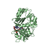
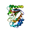
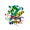
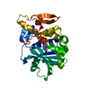
- PDB-7c6d: Crystal structure of E19A mutant chitosanase from Bacillus subtil... -
+ Open data

- Basic information
| Entry | Database: PDB / ID: 7c6d | ||||||
|---|---|---|---|---|---|---|---|
| Title | Crystal structure of E19A mutant chitosanase from Bacillus subtilis MY002 complexed with 6 GlcN. | ||||||
 Components Components | Chitosanase | ||||||
 Keywords Keywords | HYDROLASE / chitosanase | ||||||
| Function / homology |  Function and homology information Function and homology informationchitosanase / chitosanase activity / carbohydrate metabolic process / extracellular region Similarity search - Function | ||||||
| Biological species |  | ||||||
| Method |  X-RAY DIFFRACTION / SYNCHROTRON / MOLECULAR REPLACEMENT / Resolution: 1.451 Å X-RAY DIFFRACTION / SYNCHROTRON / MOLECULAR REPLACEMENT / Resolution: 1.451 Å | ||||||
 Authors Authors | Gou, Y. / Liu, Z.C. / Xie, T. / Wang, G.G. | ||||||
 Citation Citation | Journal: Colloids Surf B Biointerfaces / Year: 2021 Title: Structure-based rational design of chitosanase CsnMY002 for high yields of chitobiose. Authors: Li, Y. / Gou, Y. / Liu, Z. / Xie, T. / Wang, G. | ||||||
| History |
|
- Structure visualization
| Structure viewer | Molecule: MolmilJmol/JSmol |
|---|
- Downloads & links
-Download
| PDBx/mmCIF format | 7c6d.cif.gz | 120.2 KB | Display | PDBx/mmCIF format |
|---|---|---|---|---|
| PDB format | pdb7c6d.ent.gz | 91.2 KB | Display | PDB format |
| PDBx/mmJSON format | 7c6d.json.gz | Tree view | PDBx/mmJSON format | |
| Others |  Other downloads Other downloads |
-Validation report
| Arichive directory | https://data.pdbj.org/pub/pdb/validation_reports/c6/7c6dftp://data.pdbj.org/pub/pdb/validation_reports/c6/7c6d | HTTPS FTP |
|---|
-Related structure data
| Related structure data |  7c6cSC S: Starting model for refinement C: citing same article ( |
|---|---|
| Similar structure data |


-Links
 PDBj
PDBj- Assembly
| Deposited unit | 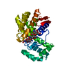
| ||||||||
|---|---|---|---|---|---|---|---|---|---|
| 1 |
| ||||||||
| Unit cell |
|
-Components
| #1: Protein | Mass: 27405.547 Da / Num. of mol.: 1 / Mutation: E19A Source method: isolated from a genetically manipulated source Source: (gene. exp.) Strain: 168 / Gene: csn, BSU26890 / Production host: |
|---|---|
| #2: Polysaccharide | 2-amino-2-deoxy-beta-D-glucopyranose-(1-4)-2-amino-2-deoxy-beta-D-glucopyranose-(1-4)-2-amino-2- ...2-amino-2-deoxy-beta-D-glucopyranose-(1-4)-2-amino-2-deoxy-beta-D-glucopyranose-(1-4)-2-amino-2-deoxy-beta-D-glucopyranose-(1-4)-2-amino-2-deoxy-beta-D-glucopyranose-(1-4)-2-amino-2-deoxy-beta-D-glucopyranose-(1-4)-2-amino-2-deoxy-beta-D-glucopyranose Source method: isolated from a genetically manipulated source |
| #3: Water | ChemComp-HOH /  Mass: 18.015 Da / Num. of mol.: 320 / Source method: isolated from a natural source / Formula: H2O Mass: 18.015 Da / Num. of mol.: 320 / Source method: isolated from a natural source / Formula: H2O |
| Has ligand of interest | N |
-Experimental details
-Experiment
| Experiment | Method: X-RAY DIFFRACTION / Number of used crystals: 1 |
|---|
- Sample preparation
| Crystal | Density Matthews: 2.64 Å3/Da / Density % sol: 53.42 % |
|---|---|
| Crystal grow | Temperature: 291 K / Method: vapor diffusion, hanging drop / pH: 7.5 Details: 0.1M HEPES pH7.5, 0.2M ammonium sulfate and 25%(w/v) PEG 3350 |
-Data collection
| Diffraction | Mean temperature: 100 K / Serial crystal experiment: N |
|---|---|
| Diffraction source | Source: SYNCHROTRON / Site: SSRF  / Beamline: BL18U1 / Wavelength: 0.97853 Å / Beamline: BL18U1 / Wavelength: 0.97853 Å |
| Detector | Type: DECTRIS PILATUS3 6M / Detector: PIXEL / Date: Nov 23, 2019 |
| Radiation | Protocol: SINGLE WAVELENGTH / Monochromatic (M) / Laue (L): M / Scattering type: x-ray |
| Radiation wavelength | Wavelength: 0.97853 Å / Relative weight: 1 |
| Reflection | Resolution: 1.45→24.98 Å / Num. obs: 51501 / % possible obs: 98.7 % / Redundancy: 12 % / Biso Wilson estimate: 12.7 Å2 / CC1/2: 0.999 / Rmerge(I) obs: 0.052 / Net I/σ(I): 47.2 |
| Reflection shell | Resolution: 1.45→1.48 Å / Rmerge(I) obs: 0.133 / Mean I/σ(I) obs: 0.147 / Num. unique obs: 2256 / CC1/2: 0.986 |
- Processing
| Software |
| ||||||||||||||||||||||||||||||||||||||||||||||||||||||||||||||||||||||||||||||||||||||||||||||||||||||||||||
|---|---|---|---|---|---|---|---|---|---|---|---|---|---|---|---|---|---|---|---|---|---|---|---|---|---|---|---|---|---|---|---|---|---|---|---|---|---|---|---|---|---|---|---|---|---|---|---|---|---|---|---|---|---|---|---|---|---|---|---|---|---|---|---|---|---|---|---|---|---|---|---|---|---|---|---|---|---|---|---|---|---|---|---|---|---|---|---|---|---|---|---|---|---|---|---|---|---|---|---|---|---|---|---|---|---|---|---|---|---|
| Refinement | Method to determine structure: MOLECULAR REPLACEMENT Starting model: 7C6C Resolution: 1.451→24.978 Å / SU ML: 0.09 / Cross valid method: THROUGHOUT / σ(F): 1.39 / Phase error: 13.42 / Stereochemistry target values: ML
| ||||||||||||||||||||||||||||||||||||||||||||||||||||||||||||||||||||||||||||||||||||||||||||||||||||||||||||
| Solvent computation | Shrinkage radii: 0.9 Å / VDW probe radii: 1.11 Å / Solvent model: FLAT BULK SOLVENT MODEL | ||||||||||||||||||||||||||||||||||||||||||||||||||||||||||||||||||||||||||||||||||||||||||||||||||||||||||||
| Displacement parameters | Biso max: 46.84 Å2 / Biso mean: 16.2764 Å2 / Biso min: 8.12 Å2 | ||||||||||||||||||||||||||||||||||||||||||||||||||||||||||||||||||||||||||||||||||||||||||||||||||||||||||||
| Refinement step | Cycle: final / Resolution: 1.451→24.978 Å
| ||||||||||||||||||||||||||||||||||||||||||||||||||||||||||||||||||||||||||||||||||||||||||||||||||||||||||||
| Refine LS restraints |
| ||||||||||||||||||||||||||||||||||||||||||||||||||||||||||||||||||||||||||||||||||||||||||||||||||||||||||||
| LS refinement shell | Refine-ID: X-RAY DIFFRACTION / Rfactor Rfree error: 0
|
