 Movie
Movie Controller

[English] 日本語
 Yorodumi
- PDB-7c38: Crystal structure of AofleA from Arthrobotrys oligospora in compl... -
+ Open data

- Basic information
| Entry | Database: PDB / ID: 7c38 | |||||||||
|---|---|---|---|---|---|---|---|---|---|---|
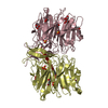
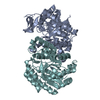
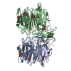
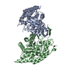
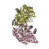
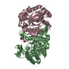
| Title | Crystal structure of AofleA from Arthrobotrys oligospora in complex with L-fucose | |||||||||
 Components Components | AofleA | |||||||||
 Keywords Keywords | SUGAR BINDING PROTEIN / nematode-trapping fungi / Arthrobotrys oligospora / Fucose-specific Lectin | |||||||||
| Function / homology | Fucose-specific lectin / Fucose-specific lectin / Fungal fucose-specific lectin / 6 Propeller / Neuraminidase / Mainly Beta / alpha-L-fucopyranose / beta-L-fucopyranose / Uncharacterized protein Function and homology information Function and homology information | |||||||||
| Biological species | Arthrobotrys oligospora | |||||||||
| Method |  X-RAY DIFFRACTION / SYNCHROTRON / MOLECULAR REPLACEMENT / Resolution: 1.2 Å X-RAY DIFFRACTION / SYNCHROTRON / MOLECULAR REPLACEMENT / Resolution: 1.2 Å | |||||||||
 Authors Authors | Liu, M. / Cheng, X. / Wang, J. / Zhang, M. / Wang, M. | |||||||||
| Funding support |  China, 2items China, 2items
| |||||||||
 Citation Citation | Journal: Int.J.Biol.Macromol. / Year: 2020 Title: Structural insights into the fungi-nematodes interaction mediated by fucose-specific lectin AofleA from Arthrobotrys oligospora. Authors: Liu, M. / Cheng, X. / Wang, J. / Tian, D. / Tang, K. / Xu, T. / Zhang, M. / Wang, Y. / Wang, M. | |||||||||
| History |
|
- Structure visualization
| Structure viewer | Molecule: MolmilJmol/JSmol |
|---|
- Downloads & links
-Download
| PDBx/mmCIF format | 7c38.cif.gz | 346.3 KB | Display | PDBx/mmCIF format |
|---|---|---|---|---|
| PDB format | pdb7c38.ent.gz | 279.8 KB | Display | PDB format |
| PDBx/mmJSON format | 7c38.json.gz | Tree view | PDBx/mmJSON format | |
| Others |  Other downloads Other downloads |
-Validation report
| Arichive directory | https://data.pdbj.org/pub/pdb/validation_reports/c3/7c38ftp://data.pdbj.org/pub/pdb/validation_reports/c3/7c38 | HTTPS FTP |
|---|
-Related structure data
| Related structure data | 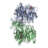 7c37SC 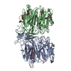 7c39C 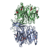 7c3cC  7c3dC 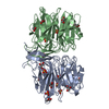 7c3eC S: Starting model for refinement C: citing same article ( |
|---|---|
| Similar structure data |


-Links
 PDBj
PDBj
- Assembly
| Deposited unit | 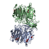
| ||||||||
|---|---|---|---|---|---|---|---|---|---|
| 1 |
| ||||||||
| Unit cell |
|
-Components
| #1: Protein | Mass: 39472.242 Da / Num. of mol.: 2 Source method: isolated from a genetically manipulated source Source: (gene. exp.)  Arthrobotrys oligospora (strain ATCC 24927 / CBS 115.81 / DSM 1491) (fungus) Arthrobotrys oligospora (strain ATCC 24927 / CBS 115.81 / DSM 1491) (fungus)Strain: ATCC 24927 / CBS 115.81 / DSM 1491 / Gene: AOL_s00076g540 / Plasmid: pET28a / Production host:  #2: Sugar | ChemComp-FUC /   Type: L-saccharide, alpha linking / Mass: 164.156 Da / Num. of mol.: 4 Type: L-saccharide, alpha linking / Mass: 164.156 Da / Num. of mol.: 4Source method: isolated from a genetically manipulated source Formula: C6H12O5 / Feature type: SUBJECT OF INVESTIGATION #3: Sugar | ChemComp-FUL / 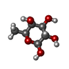  Type: L-saccharide, beta linking / Mass: 164.156 Da / Num. of mol.: 4 Type: L-saccharide, beta linking / Mass: 164.156 Da / Num. of mol.: 4Source method: isolated from a genetically manipulated source Formula: C6H12O5 / Feature type: SUBJECT OF INVESTIGATION #4: Chemical | ChemComp-GOL /   Mass: 92.094 Da / Num. of mol.: 4 / Source method: obtained synthetically / Formula: C3H8O3 Mass: 92.094 Da / Num. of mol.: 4 / Source method: obtained synthetically / Formula: C3H8O3#5: Water | ChemComp-HOH / |  Mass: 18.015 Da / Num. of mol.: 1260 / Source method: isolated from a natural source / Formula: H2O Mass: 18.015 Da / Num. of mol.: 1260 / Source method: isolated from a natural source / Formula: H2OHas ligand of interest | Y | |
|---|
-Experimental details
-Experiment
| Experiment | Method: X-RAY DIFFRACTION / Number of used crystals: 1 |
|---|
- Sample preparation
| Crystal | Density Matthews: 2.24 Å3/Da / Density % sol: 45.14 % |
|---|---|
| Crystal grow | Temperature: 289 K / Method: vapor diffusion, sitting drop / pH: 5.5 Details: 0.1 M Ssodium citrate pH 5.5 and 22% (w/v) PEG 1000 |
-Data collection
| Diffraction | Mean temperature: 100 K / Serial crystal experiment: N | |||||||||||||||||||||||||||||||||||||||||||||||||||||||||||||||||||||||||||||||||||||||||||||||||||
|---|---|---|---|---|---|---|---|---|---|---|---|---|---|---|---|---|---|---|---|---|---|---|---|---|---|---|---|---|---|---|---|---|---|---|---|---|---|---|---|---|---|---|---|---|---|---|---|---|---|---|---|---|---|---|---|---|---|---|---|---|---|---|---|---|---|---|---|---|---|---|---|---|---|---|---|---|---|---|---|---|---|---|---|---|---|---|---|---|---|---|---|---|---|---|---|---|---|---|---|---|
| Diffraction source | Source: SYNCHROTRON / Site: SSRF / Beamline: BL17U / Wavelength: 0.9792 Å | |||||||||||||||||||||||||||||||||||||||||||||||||||||||||||||||||||||||||||||||||||||||||||||||||||
| Detector | Type: DECTRIS EIGER X 16M / Detector: PIXEL / Date: Mar 23, 2018 | |||||||||||||||||||||||||||||||||||||||||||||||||||||||||||||||||||||||||||||||||||||||||||||||||||
| Radiation | Protocol: SINGLE WAVELENGTH / Monochromatic (M) / Laue (L): M / Scattering type: x-ray | |||||||||||||||||||||||||||||||||||||||||||||||||||||||||||||||||||||||||||||||||||||||||||||||||||
| Radiation wavelength | Wavelength: 0.9792 Å / Relative weight: 1 | |||||||||||||||||||||||||||||||||||||||||||||||||||||||||||||||||||||||||||||||||||||||||||||||||||
| Reflection | Resolution: 1.2→50 Å / Num. obs: 210395 / % possible obs: 94.7 % / Redundancy: 5.8 % / Rmerge(I) obs: 0.068 / Rpim(I) all: 0.03 / Rrim(I) all: 0.075 / Χ2: 1.014 / Net I/σ(I): 14.9 / Num. measured all: 1226269 | |||||||||||||||||||||||||||||||||||||||||||||||||||||||||||||||||||||||||||||||||||||||||||||||||||
| Reflection shell | Diffraction-ID: 1
|
- Processing
| Software |
| ||||||||||||||||||||||||||||||||||||||||||||||||||||||||||||||||||||||||||||||||||||||||||||||||||||||||||||||||||||||||||||||||||||||||||||||||||||||||||||||||||||||||||||||||||||||||||
|---|---|---|---|---|---|---|---|---|---|---|---|---|---|---|---|---|---|---|---|---|---|---|---|---|---|---|---|---|---|---|---|---|---|---|---|---|---|---|---|---|---|---|---|---|---|---|---|---|---|---|---|---|---|---|---|---|---|---|---|---|---|---|---|---|---|---|---|---|---|---|---|---|---|---|---|---|---|---|---|---|---|---|---|---|---|---|---|---|---|---|---|---|---|---|---|---|---|---|---|---|---|---|---|---|---|---|---|---|---|---|---|---|---|---|---|---|---|---|---|---|---|---|---|---|---|---|---|---|---|---|---|---|---|---|---|---|---|---|---|---|---|---|---|---|---|---|---|---|---|---|---|---|---|---|---|---|---|---|---|---|---|---|---|---|---|---|---|---|---|---|---|---|---|---|---|---|---|---|---|---|---|---|---|---|---|---|---|
| Refinement | Method to determine structure: MOLECULAR REPLACEMENT Starting model: 7C37 Resolution: 1.2→27.349 Å / SU ML: 0.07 / Cross valid method: THROUGHOUT / σ(F): 1.35 / Phase error: 11.17 / Stereochemistry target values: ML
| ||||||||||||||||||||||||||||||||||||||||||||||||||||||||||||||||||||||||||||||||||||||||||||||||||||||||||||||||||||||||||||||||||||||||||||||||||||||||||||||||||||||||||||||||||||||||||
| Solvent computation | Shrinkage radii: 0.9 Å / VDW probe radii: 1.11 Å / Solvent model: FLAT BULK SOLVENT MODEL | ||||||||||||||||||||||||||||||||||||||||||||||||||||||||||||||||||||||||||||||||||||||||||||||||||||||||||||||||||||||||||||||||||||||||||||||||||||||||||||||||||||||||||||||||||||||||||
| Displacement parameters | Biso max: 65.5 Å2 / Biso mean: 14.1833 Å2 / Biso min: 4.56 Å2 | ||||||||||||||||||||||||||||||||||||||||||||||||||||||||||||||||||||||||||||||||||||||||||||||||||||||||||||||||||||||||||||||||||||||||||||||||||||||||||||||||||||||||||||||||||||||||||
| Refinement step | Cycle: final / Resolution: 1.2→27.349 Å
| ||||||||||||||||||||||||||||||||||||||||||||||||||||||||||||||||||||||||||||||||||||||||||||||||||||||||||||||||||||||||||||||||||||||||||||||||||||||||||||||||||||||||||||||||||||||||||
| LS refinement shell | Refine-ID: X-RAY DIFFRACTION / Rfactor Rfree error: 0
|
