 Movie
Movie Controller


+ Open data

- Basic information
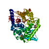
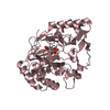
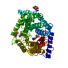
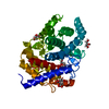
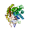
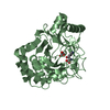
| Entry | Database: PDB / ID: 7c24 | ||||||
|---|---|---|---|---|---|---|---|
| Title | Glycosidase F290Y at pH8.0 | ||||||
 Components Components | Isomaltose glucohydrolase | ||||||
 Keywords Keywords | HYDROLASE / Glycoside hydrolase | ||||||
| Function / homology |  Function and homology information Function and homology informationisomaltose glucohydrolase / polysaccharide catabolic process / hydrolase activity, hydrolyzing O-glycosyl compounds / cytoplasm Similarity search - Function | ||||||
| Biological species |  Kribbella flavida (bacteria) Kribbella flavida (bacteria) | ||||||
| Method |  X-RAY DIFFRACTION / SYNCHROTRON / MOLECULAR REPLACEMENT / Resolution: 1.71 Å X-RAY DIFFRACTION / SYNCHROTRON / MOLECULAR REPLACEMENT / Resolution: 1.71 Å | ||||||
 Authors Authors | Tagami, T. / Kikuchi, A. / Okuyama, M. / Kimura, A. | ||||||
 Citation Citation | Journal: Febs J. / Year: 2022 Title: Structural insights reveal the second base catalyst of isomaltose glucohydrolase. Authors: Tagami, T. / Chen, M. / Furunaga, Y. / Kikuchi, A. / Sadahiro, J. / Lang, W. / Okuyama, M. / Tanaka, Y. / Iwasaki, T. / Yao, M. / Kimura, A. | ||||||
| History |
|
- Structure visualization
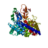
| Structure viewer | Molecule: MolmilJmol/JSmol |
|---|
- Downloads & links
-Download
| PDBx/mmCIF format | 7c24.cif.gz | 100.5 KB | Display | PDBx/mmCIF format |
|---|---|---|---|---|
| PDB format | pdb7c24.ent.gz | 70.8 KB | Display | PDB format |
| PDBx/mmJSON format | 7c24.json.gz | Tree view | PDBx/mmJSON format | |
| Others |  Other downloads Other downloads |
-Validation report
| Arichive directory | https://data.pdbj.org/pub/pdb/validation_reports/c2/7c24ftp://data.pdbj.org/pub/pdb/validation_reports/c2/7c24 | HTTPS FTP |
|---|
-Related structure data
| Related structure data |  5z3aC  5z3bC  5z3cC  5z3dSC 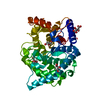 5z3eC 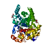 5z3fC  7c25C  7c26C  7c27C S: Starting model for refinement C: citing same article ( |
|---|---|
| Similar structure data |


-Links
 PDBj
PDBj

- Assembly
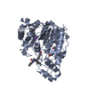
| Deposited unit | 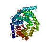
| |||||||||||||||
|---|---|---|---|---|---|---|---|---|---|---|---|---|---|---|---|---|
| 1 |
| |||||||||||||||
| Unit cell |
| |||||||||||||||
| Components on special symmetry positions |
|
-Components
| #1: Protein | Mass: 44060.059 Da / Num. of mol.: 1 / Mutation: F290Y Source method: isolated from a genetically manipulated source Source: (gene. exp.) Kribbella flavida (strain DSM 17836 / JCM 10339 / NBRC 14399) (bacteria)Strain: DSM 17836 / JCM 10339 / NBRC 14399 / Gene: Kfla_1896 / Production host: | ||||||
|---|---|---|---|---|---|---|---|
| #2: Chemical | ChemComp-GOL /   Mass: 92.094 Da / Num. of mol.: 5 / Source method: obtained synthetically / Formula: C3H8O3 Mass: 92.094 Da / Num. of mol.: 5 / Source method: obtained synthetically / Formula: C3H8O3#3: Chemical | ChemComp-NH4 / |   Mass: 18.038 Da / Num. of mol.: 1 / Source method: obtained synthetically / Formula: H4N Mass: 18.038 Da / Num. of mol.: 1 / Source method: obtained synthetically / Formula: H4N#4: Water | ChemComp-HOH / |  Mass: 18.015 Da / Num. of mol.: 349 / Source method: isolated from a natural source / Formula: H2O Mass: 18.015 Da / Num. of mol.: 349 / Source method: isolated from a natural source / Formula: H2OHas ligand of interest | N | |
-Experimental details
-Experiment
| Experiment | Method: X-RAY DIFFRACTION / Number of used crystals: 1 |
|---|
- Sample preparation
| Crystal | Density Matthews: 2.8 Å3/Da / Density % sol: 56.06 % |
|---|---|
| Crystal grow | Temperature: 293 K / Method: vapor diffusion, hanging drop / pH: 8 Details: 0.2 M Ammonium citrate (pH 8.0), 8% PEG monomethyl ether 2000 |
-Data collection
| Diffraction | Mean temperature: 100 K / Serial crystal experiment: N |
|---|---|
| Diffraction source | Source: SYNCHROTRON / Site: Photon Factory  / Beamline: AR-NW12A / Wavelength: 1 Å / Beamline: AR-NW12A / Wavelength: 1 Å |
| Detector | Type: DECTRIS PILATUS3 S 2M / Detector: PIXEL / Date: Dec 6, 2019 |
| Radiation | Protocol: SINGLE WAVELENGTH / Monochromatic (M) / Laue (L): M / Scattering type: x-ray |
| Radiation wavelength | Wavelength: 1 Å / Relative weight: 1 |
| Reflection | Resolution: 1.71→41.59 Å / Num. obs: 54864 / % possible obs: 99.9 % / Redundancy: 26.1 % / CC1/2: 0.999 / Rrim(I) all: 0.226 / Rsym value: 0.222 / Net I/σ(I): 0.134 |
| Reflection shell | Resolution: 1.71→1.81 Å / Redundancy: 25.5 % / Mean I/σ(I) obs: 0.0084 / Num. unique obs: 8723 / CC1/2: 0.529 / Rrim(I) all: 4.667 / Rsym value: 4.575 / % possible all: 99.7 |
- Processing
| Software |
| |||||||||||||||||||||||||||||||||||||||||||||||||||||||||||||||||||||||||||||||||||||||||||||||||||||||||||||||||||||||||||||||||||||||||||||||||||||||||||
|---|---|---|---|---|---|---|---|---|---|---|---|---|---|---|---|---|---|---|---|---|---|---|---|---|---|---|---|---|---|---|---|---|---|---|---|---|---|---|---|---|---|---|---|---|---|---|---|---|---|---|---|---|---|---|---|---|---|---|---|---|---|---|---|---|---|---|---|---|---|---|---|---|---|---|---|---|---|---|---|---|---|---|---|---|---|---|---|---|---|---|---|---|---|---|---|---|---|---|---|---|---|---|---|---|---|---|---|---|---|---|---|---|---|---|---|---|---|---|---|---|---|---|---|---|---|---|---|---|---|---|---|---|---|---|---|---|---|---|---|---|---|---|---|---|---|---|---|---|---|---|---|---|---|---|---|---|
| Refinement | Method to determine structure: MOLECULAR REPLACEMENT Starting model: 5Z3D Resolution: 1.71→41.59 Å / Cor.coef. Fo:Fc: 0.977 / Cor.coef. Fo:Fc free: 0.969 / SU B: 2.562 / SU ML: 0.075 / Cross valid method: FREE R-VALUE / ESU R: 0.082 / ESU R Free: 0.083 Details: Hydrogens have been added in their riding positions
| |||||||||||||||||||||||||||||||||||||||||||||||||||||||||||||||||||||||||||||||||||||||||||||||||||||||||||||||||||||||||||||||||||||||||||||||||||||||||||
| Solvent computation | Ion probe radii: 0.8 Å / Shrinkage radii: 0.8 Å / VDW probe radii: 1.2 Å | |||||||||||||||||||||||||||||||||||||||||||||||||||||||||||||||||||||||||||||||||||||||||||||||||||||||||||||||||||||||||||||||||||||||||||||||||||||||||||
| Displacement parameters | Biso mean: 27.482 Å2
| |||||||||||||||||||||||||||||||||||||||||||||||||||||||||||||||||||||||||||||||||||||||||||||||||||||||||||||||||||||||||||||||||||||||||||||||||||||||||||
| Refinement step | Cycle: LAST / Resolution: 1.71→41.59 Å
| |||||||||||||||||||||||||||||||||||||||||||||||||||||||||||||||||||||||||||||||||||||||||||||||||||||||||||||||||||||||||||||||||||||||||||||||||||||||||||
| Refine LS restraints |
| |||||||||||||||||||||||||||||||||||||||||||||||||||||||||||||||||||||||||||||||||||||||||||||||||||||||||||||||||||||||||||||||||||||||||||||||||||||||||||
| LS refinement shell |
|
