 Movie
Movie Controller

[English] 日本語
 Yorodumi
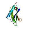
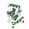
- PDB-7c21: Crystal structure of Duvenhage virus phosphoprotein C-terminal domain -
+ Open data

- Basic information
| Entry | Database: PDB / ID: 7c21 | ||||||
|---|---|---|---|---|---|---|---|
| Title | Crystal structure of Duvenhage virus phosphoprotein C-terminal domain | ||||||
 Components Components | Phosphoprotein | ||||||
 Keywords Keywords | VIRAL PROTEIN / phosphoprotein | ||||||
| Function / homology |  Function and homology information Function and homology information: / viral transcription / symbiont-mediated suppression of host JAK-STAT cascade via inhibition of STAT2 activity / symbiont-mediated suppression of host JAK-STAT cascade via inhibition of STAT1 activity / virion component / host cell cytoplasm / symbiont-mediated suppression of host innate immune response / symbiont-mediated suppression of host type I interferon-mediated signaling pathway / RNA-directed RNA polymerase activity / cytoplasm Similarity search - Function | ||||||
| Biological species |  Duvenhage virus Duvenhage virus | ||||||
| Method |  X-RAY DIFFRACTION / SYNCHROTRON / MOLECULAR REPLACEMENT / Resolution: 1.95 Å X-RAY DIFFRACTION / SYNCHROTRON / MOLECULAR REPLACEMENT / Resolution: 1.95 Å | ||||||
 Authors Authors | Sugiyama, A. / Jiang, X. / Maenaka, K. / Yao, M. / Ose, T. | ||||||
| Funding support |  Japan, 1items Japan, 1items
| ||||||
 Citation Citation | Journal: Biochem.Biophys.Res.Commun. / Year: 2020 Title: Structural comparison of the C-terminal domain of functionally divergent lyssavirus P proteins. Authors: Sugiyama, A. / Nomai, T. / Jiang, X. / Minami, M. / Yao, M. / Maenaka, K. / Ito, N. / Gooley, P.R. / Moseley, G.W. / Ose, T. | ||||||
| History |
|
- Structure visualization
| Structure viewer | Molecule: MolmilJmol/JSmol |
|---|
- Downloads & links
-Download
| PDBx/mmCIF format | 7c21.cif.gz | 35.7 KB | Display | PDBx/mmCIF format |
|---|---|---|---|---|
| PDB format | pdb7c21.ent.gz | 22.2 KB | Display | PDB format |
| PDBx/mmJSON format | 7c21.json.gz | Tree view | PDBx/mmJSON format | |
| Others |  Other downloads Other downloads |
-Validation report
| Arichive directory | https://data.pdbj.org/pub/pdb/validation_reports/c2/7c21ftp://data.pdbj.org/pub/pdb/validation_reports/c2/7c21 | HTTPS FTP |
|---|
-Related structure data
| Related structure data | 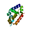 7c20C  1vyiS S: Starting model for refinement C: citing same article ( |
|---|---|
| Similar structure data |


-Links
 PDBj
PDBj- Assembly
| Deposited unit | 
| ||||||||
|---|---|---|---|---|---|---|---|---|---|
| 1 |
| ||||||||
| Unit cell |
|
-Components
| #1: Protein | Mass: 12870.816 Da / Num. of mol.: 1 Source method: isolated from a genetically manipulated source Source: (gene. exp.) Duvenhage virus / Production host:  |
|---|---|
| #2: Water | ChemComp-HOH /  Mass: 18.015 Da / Num. of mol.: 52 / Source method: isolated from a natural source / Formula: H2O Mass: 18.015 Da / Num. of mol.: 52 / Source method: isolated from a natural source / Formula: H2O |
-Experimental details
-Experiment
| Experiment | Method: X-RAY DIFFRACTION / Number of used crystals: 1 |
|---|
- Sample preparation
| Crystal | Density Matthews: 2.42 Å3/Da / Density % sol: 49.11 % |
|---|---|
| Crystal grow | Temperature: 293 K / Method: vapor diffusion, sitting drop / Details: 0.1 M HEPES pH7.5, 20% PEG 8000 |
-Data collection
| Diffraction | Mean temperature: 100 K / Serial crystal experiment: N |
|---|---|
| Diffraction source | Source: SYNCHROTRON / Site: SPring-8 / Beamline: BL45XU / Wavelength: 1 Å |
| Detector | Type: DECTRIS PILATUS3 S 6M / Detector: PIXEL / Date: Nov 30, 2019 |
| Radiation | Protocol: SINGLE WAVELENGTH / Monochromatic (M) / Laue (L): M / Scattering type: x-ray |
| Radiation wavelength | Wavelength: 1 Å / Relative weight: 1 |
| Reflection | Resolution: 1.95→43.8 Å / Num. obs: 8380 / % possible obs: 98 % / Redundancy: 8.6 % / Biso Wilson estimate: 23.57 Å2 / CC1/2: 0.99 / Rrim(I) all: 0.216 / Net I/σ(I): 7.53 |
| Reflection shell | Resolution: 1.95→2.07 Å / Redundancy: 8.6 % / Mean I/σ(I) obs: 1.56 / Num. unique obs: 1361 / CC1/2: 0.607 / Rrim(I) all: 1.385 / % possible all: 97.4 |
- Processing
| Software |
| |||||||||||||||||||||||||||||||||||
|---|---|---|---|---|---|---|---|---|---|---|---|---|---|---|---|---|---|---|---|---|---|---|---|---|---|---|---|---|---|---|---|---|---|---|---|---|
| Refinement | Method to determine structure: MOLECULAR REPLACEMENT Starting model: 1VYI Resolution: 1.95→43.8 Å / SU ML: 0.26 / Cross valid method: FREE R-VALUE / σ(F): 1.34 / Phase error: 27.09
| |||||||||||||||||||||||||||||||||||
| Solvent computation | Shrinkage radii: 0.9 Å / VDW probe radii: 1.11 Å | |||||||||||||||||||||||||||||||||||
| Refinement step | Cycle: LAST / Resolution: 1.95→43.8 Å
| |||||||||||||||||||||||||||||||||||
| Refine LS restraints |
| |||||||||||||||||||||||||||||||||||
| LS refinement shell |
|
