 Movie
Movie Controller


+ Open data

- Basic information
| Entry | Database: PDB / ID: 7c0g | ||||||
|---|---|---|---|---|---|---|---|
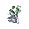
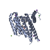
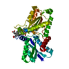
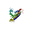
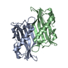
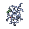
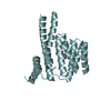
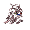
| Title | Aca1 in complex with 14bp palindromic DNA target | ||||||
 Components Components |
| ||||||
 Keywords Keywords | TRANSCRIPTION / CRISPR / anti-CRISPR / anti-CRISPR-associated / DNA binding / autoregulation / Aca1 / complex | ||||||
| Function / homology | Lambda repressor-like, DNA-binding domain superfamily / DNA binding / DNA / DNA (> 10) / HTH cro/C1-type domain-containing protein Function and homology information Function and homology information | ||||||
| Biological species |  Pseudomonas phage JBD30 (virus) Pseudomonas phage JBD30 (virus)synthetic construct (others) | ||||||
| Method |  X-RAY DIFFRACTION / SYNCHROTRON / MOLECULAR REPLACEMENT / Resolution: 2.4 Å X-RAY DIFFRACTION / SYNCHROTRON / MOLECULAR REPLACEMENT / Resolution: 2.4 Å | ||||||
 Authors Authors | Liu, Y.H. / Zhang, L.S. / Wu, B.X. / Huang, H.D. | ||||||
 Citation Citation | Journal: To Be Published Title: Aca1 in complex with 14bp palindromic DNA target Authors: Liu, Y.H. / Zhang, L.S. / Wu, B.X. / Huang, H.D. | ||||||
| History |
|
- Structure visualization
| Structure viewer | Molecule: MolmilJmol/JSmol |
|---|
- Downloads & links
-Download
| PDBx/mmCIF format | 7c0g.cif.gz | 58.7 KB | Display | PDBx/mmCIF format |
|---|---|---|---|---|
| PDB format | pdb7c0g.ent.gz | 40.8 KB | Display | PDB format |
| PDBx/mmJSON format | 7c0g.json.gz | Tree view | PDBx/mmJSON format | |
| Others |  Other downloads Other downloads |
-Validation report
| Summary document | 7c0g_validation.pdf.gz | 445.2 KB | Display | wwPDB validaton report |
|---|---|---|---|---|
| Full document | 7c0g_full_validation.pdf.gz | 448.5 KB | Display | |
| Data in XML | 7c0g_validation.xml.gz | 9.4 KB | Display | |
| Data in CIF | 7c0g_validation.cif.gz | 12.7 KB | Display | |
| Arichive directory | https://data.pdbj.org/pub/pdb/validation_reports/c0/7c0gftp://data.pdbj.org/pub/pdb/validation_reports/c0/7c0g | HTTPS FTP |
-Related structure data
| Related structure data |  7c0b 7c0a S: Starting model for refinement |
|---|---|
| Similar structure data |


-Links
 PDBj
PDBj


- Assembly
| Deposited unit | 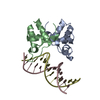
| ||||||||||||
|---|---|---|---|---|---|---|---|---|---|---|---|---|---|
| 1 |
| ||||||||||||
| Unit cell |
|
-Components
| #1: Protein | Mass: 8908.148 Da / Num. of mol.: 2 Source method: isolated from a genetically manipulated source Source: (gene. exp.) Pseudomonas phage JBD30 (virus) / Gene: JBD30_036 / Production host:  #2: DNA chain | Mass: 4281.779 Da / Num. of mol.: 2 / Source method: obtained synthetically / Source: (synth.) synthetic construct (others) #3: Water | ChemComp-HOH / |  Mass: 18.015 Da / Num. of mol.: 86 / Source method: isolated from a natural source / Formula: H2O Mass: 18.015 Da / Num. of mol.: 86 / Source method: isolated from a natural source / Formula: H2O |
|---|
-Experimental details
-Experiment
| Experiment | Method: X-RAY DIFFRACTION / Number of used crystals: 1 |
|---|
- Sample preparation
| Crystal | Density Matthews: 2.65 Å3/Da / Density % sol: 53.57 % |
|---|---|
| Crystal grow | Temperature: 293 K / Method: vapor diffusion, sitting drop / Details: 0.1M Sodium acetate pH5.0; 20%(w/v) MPD |
-Data collection
| Diffraction | Mean temperature: 100 K / Serial crystal experiment: N |
|---|---|
| Diffraction source | Source: SYNCHROTRON / Site: SSRF  / Beamline: BL19U1 / Wavelength: 0.9789 Å / Beamline: BL19U1 / Wavelength: 0.9789 Å |
| Detector | Type: DECTRIS PILATUS3 S 6M / Detector: PIXEL / Date: Oct 15, 2019 |
| Radiation | Protocol: SINGLE WAVELENGTH / Monochromatic (M) / Laue (L): M / Scattering type: x-ray |
| Radiation wavelength | Wavelength: 0.9789 Å / Relative weight: 1 |
| Reflection | Resolution: 2.4→40 Å / Num. obs: 12045 / % possible obs: 99.5 % / Redundancy: 27.4 % / CC1/2: 1 / CC star: 1 / Rmerge(I) obs: 0.133 / Rsym value: 0.133 / Net I/σ(I): 24.333 |
| Reflection shell | Resolution: 2.4→2.49 Å / Rmerge(I) obs: 0.814 / Num. unique obs: 1108 / CC1/2: 0.385 |
- Processing
| Software |
| ||||||||||||||||||||||||
|---|---|---|---|---|---|---|---|---|---|---|---|---|---|---|---|---|---|---|---|---|---|---|---|---|---|
| Refinement | Method to determine structure: MOLECULAR REPLACEMENT Starting model: 7C0B 7c0b Resolution: 2.4→35 Å / Cross valid method: FREE R-VALUE
| ||||||||||||||||||||||||
| Displacement parameters | Biso mean: 31.69 Å2 | ||||||||||||||||||||||||
| Refinement step | Cycle: LAST / Resolution: 2.4→35 Å
| ||||||||||||||||||||||||
| Refine LS restraints |
|
