 Movie
Movie Controller

[English] 日本語
 Yorodumi
- PDB-7c03: Crystal structure of POLArISact(T57S), genetically encoded probe ... -
+ Open data

- Basic information
| Entry | Database: PDB / ID: 7c03 | |||||||||
|---|---|---|---|---|---|---|---|---|---|---|
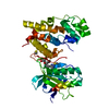
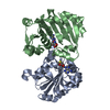
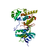
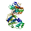
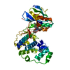
| Title | Crystal structure of POLArISact(T57S), genetically encoded probe for fluorescent polarization | |||||||||
 Components Components | POLArISact(T57S) | |||||||||
 Keywords Keywords | LUMINESCENT PROTEIN / Fluorescent protein Fluorescent polarization Protein engineering | |||||||||
| Function / homology | Nuclear Transport Factor 2; Chain: A, - #10 / Nuclear Transport Factor 2; Chain: A, / Roll / Alpha Beta Function and homology information Function and homology information | |||||||||
| Biological species | synthetic construct (others) | |||||||||
| Method |  X-RAY DIFFRACTION / SYNCHROTRON / MOLECULAR REPLACEMENT / molecular replacement / Resolution: 2.501 Å X-RAY DIFFRACTION / SYNCHROTRON / MOLECULAR REPLACEMENT / molecular replacement / Resolution: 2.501 Å | |||||||||
 Authors Authors | Tomabechi, Y. / Sakai, N. / Shirouzu, M. | |||||||||
 Citation Citation | Journal: Proc.Natl.Acad.Sci.USA / Year: 2021 Title: POLArIS, a versatile probe for molecular orientation, revealed actin filaments associated with microtubule asters in early embryos. Authors: Sugizaki, A. / Sato, K. / Chiba, K. / Saito, K. / Kawagishi, M. / Tomabechi, Y. / Mehta, S.B. / Ishii, H. / Sakai, N. / Shirouzu, M. / Tani, T. / Terada, S. | |||||||||
| History |
|
- Structure visualization
| Structure viewer | Molecule: MolmilJmol/JSmol |
|---|
- Downloads & links
-Download
| PDBx/mmCIF format | 7c03.cif.gz | 80.3 KB | Display | PDBx/mmCIF format |
|---|---|---|---|---|
| PDB format | pdb7c03.ent.gz | 58.1 KB | Display | PDB format |
| PDBx/mmJSON format | 7c03.json.gz | Tree view | PDBx/mmJSON format | |
| Others |  Other downloads Other downloads |
-Validation report
| Summary document | 7c03_validation.pdf.gz | 435.6 KB | Display | wwPDB validaton report |
|---|---|---|---|---|
| Full document | 7c03_full_validation.pdf.gz | 439.2 KB | Display | |
| Data in XML | 7c03_validation.xml.gz | 14.2 KB | Display | |
| Data in CIF | 7c03_validation.cif.gz | 18.9 KB | Display | |
| Arichive directory | https://data.pdbj.org/pub/pdb/validation_reports/c0/7c03ftp://data.pdbj.org/pub/pdb/validation_reports/c0/7c03 | HTTPS FTP |
-Related structure data
| Related structure data | 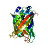 3evpS S: Starting model for refinement |
|---|---|
| Similar structure data |


-Links
 PDBj
PDBj- Assembly
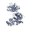
| Deposited unit | 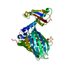
| ||||||||
|---|---|---|---|---|---|---|---|---|---|
| 1 |
| ||||||||
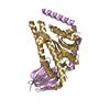
| 2 | 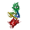
| ||||||||
| Unit cell |
|
-Components
| #1: Protein | Mass: 39081.215 Da / Num. of mol.: 1 Source method: isolated from a genetically manipulated source Source: (gene. exp.) synthetic construct (others) / Production host:  |
|---|---|
| #2: Water | ChemComp-HOH /  Mass: 18.015 Da / Num. of mol.: 81 / Source method: isolated from a natural source / Formula: H2O Mass: 18.015 Da / Num. of mol.: 81 / Source method: isolated from a natural source / Formula: H2O |
| Has ligand of interest | Y |
| Has protein modification | Y |
-Experimental details
-Experiment
| Experiment | Method: X-RAY DIFFRACTION / Number of used crystals: 1 |
|---|
- Sample preparation
| Crystal | Density Matthews: 3.76 Å3/Da / Density % sol: 67.33 % |
|---|---|
| Crystal grow | Temperature: 293 K / Method: vapor diffusion, sitting drop Details: 0.1M sodium acetate (pH4.5-5.0), 6-8% (v/v) PEG 4000 |
-Data collection
| Diffraction | Mean temperature: 100 K / Serial crystal experiment: N |
|---|---|
| Diffraction source | Source: SYNCHROTRON / Site: NSLS  / Beamline: X6A / Wavelength: 1 Å / Beamline: X6A / Wavelength: 1 Å |
| Detector | Type: ADSC QUANTUM 210 / Detector: CCD / Date: Sep 7, 2018 |
| Radiation | Protocol: SINGLE WAVELENGTH / Monochromatic (M) / Laue (L): M / Scattering type: x-ray |
| Radiation wavelength | Wavelength: 1 Å / Relative weight: 1 |
| Reflection | Resolution: 2.5→50 Å / Num. obs: 20785 / % possible obs: 99.9 % / Redundancy: 7.1 % / CC1/2: 0.912 / Net I/σ(I): 13.3 |
| Reflection shell | Resolution: 2.5→2.64 Å / Num. unique obs: 45440 / CC1/2: 0.644 |
-Phasing
| Phasing | Method: molecular replacement | |||||||||
|---|---|---|---|---|---|---|---|---|---|---|
| Phasing MR |
|
- Processing
| Software |
| |||||||||||||||||||||||||||||||||||||||||||||||||||||||||||||||||||||||||||||||||||||||||||||||||||||||||
|---|---|---|---|---|---|---|---|---|---|---|---|---|---|---|---|---|---|---|---|---|---|---|---|---|---|---|---|---|---|---|---|---|---|---|---|---|---|---|---|---|---|---|---|---|---|---|---|---|---|---|---|---|---|---|---|---|---|---|---|---|---|---|---|---|---|---|---|---|---|---|---|---|---|---|---|---|---|---|---|---|---|---|---|---|---|---|---|---|---|---|---|---|---|---|---|---|---|---|---|---|---|---|---|---|---|---|
| Refinement | Method to determine structure: MOLECULAR REPLACEMENT Starting model: 3EVP Resolution: 2.501→46.202 Å / SU ML: 0.47 / Cross valid method: THROUGHOUT / σ(F): 1.34 / Phase error: 28.61 / Stereochemistry target values: ML
| |||||||||||||||||||||||||||||||||||||||||||||||||||||||||||||||||||||||||||||||||||||||||||||||||||||||||
| Solvent computation | Shrinkage radii: 0.9 Å / VDW probe radii: 1.11 Å / Solvent model: FLAT BULK SOLVENT MODEL | |||||||||||||||||||||||||||||||||||||||||||||||||||||||||||||||||||||||||||||||||||||||||||||||||||||||||
| Displacement parameters | Biso max: 198.21 Å2 / Biso mean: 89.6744 Å2 / Biso min: 42.09 Å2 | |||||||||||||||||||||||||||||||||||||||||||||||||||||||||||||||||||||||||||||||||||||||||||||||||||||||||
| Refinement step | Cycle: final / Resolution: 2.501→46.202 Å
| |||||||||||||||||||||||||||||||||||||||||||||||||||||||||||||||||||||||||||||||||||||||||||||||||||||||||
| LS refinement shell | Refine-ID: X-RAY DIFFRACTION / Rfactor Rfree error: 0 / Total num. of bins used: 14
|
