 Movie
Movie Controller


+ Open data

- Basic information
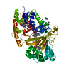
| Entry | Database: PDB / ID: 7bz0 | ||||||
|---|---|---|---|---|---|---|---|
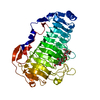
| Title | complex structure of alginate lyase AlyF-OU02 with G6 | ||||||
 Components Components | Alginate lyase AlyF-OU02 | ||||||
 Keywords Keywords | LYASE / PL6 / Ca2+-independent / complex / substrate-binding mechanism. | ||||||
| Function / homology | Single-stranded right-handed beta-helix, Pectin lyase-like / Pectate Lyase C-like / 3 Solenoid / Mainly Beta Function and homology information Function and homology information | ||||||
| Biological species |  Vibrio splendidus (bacteria) Vibrio splendidus (bacteria) | ||||||
| Method |  X-RAY DIFFRACTION / SYNCHROTRON / MOLECULAR REPLACEMENT / Resolution: 1.8 Å X-RAY DIFFRACTION / SYNCHROTRON / MOLECULAR REPLACEMENT / Resolution: 1.8 Å | ||||||
 Authors Authors | Liu, W. / Lyu, Q. / Zhang, K. | ||||||
| Funding support |  China, 1items China, 1items
| ||||||
 Citation Citation | Journal: Acta Crystallogr D Struct Biol / Year: 2021 Title: Structural insights into the substrate-binding cleft of AlyF reveal the first long-chain alginate-binding mode. Authors: Zhang, K. / Liu, T. / Liu, W. / Lyu, Q. | ||||||
| History |
|
- Structure visualization
| Structure viewer | Molecule: MolmilJmol/JSmol |
|---|
- Downloads & links
-Download
| PDBx/mmCIF format | 7bz0.cif.gz | 120.4 KB | Display | PDBx/mmCIF format |
|---|---|---|---|---|
| PDB format | pdb7bz0.ent.gz | 88.6 KB | Display | PDB format |
| PDBx/mmJSON format | 7bz0.json.gz | Tree view | PDBx/mmJSON format | |
| Others |  Other downloads Other downloads |
-Validation report
| Arichive directory | https://data.pdbj.org/pub/pdb/validation_reports/bz/7bz0ftp://data.pdbj.org/pub/pdb/validation_reports/bz/7bz0 | HTTPS FTP |
|---|
-Related structure data
| Related structure data |  6itgS S: Starting model for refinement |
|---|---|
| Similar structure data |


-Links
 PDBj
PDBj- Assembly
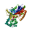
| Deposited unit | 
| ||||||||
|---|---|---|---|---|---|---|---|---|---|
| 1 |
| ||||||||
| Unit cell |
|
-Components
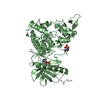
| #1: Protein | Mass: 59022.688 Da / Num. of mol.: 1 / Mutation: K272A Source method: isolated from a genetically manipulated source Details: AlyF is a Ca2+-independent alginate lyase and specially degrades polyG to produce trisaccharides as the main products. Source: (gene. exp.) Vibrio splendidus (bacteria) / Production host: References: Lyases; Carbon-oxygen lyases; Acting on polysaccharides |
|---|---|
| #2: Polysaccharide | alpha-L-gulopyranuronic acid-(1-4)-alpha-L-gulopyranuronic acid-(1-4)-alpha-L-gulopyranuronic acid- ...alpha-L-gulopyranuronic acid-(1-4)-alpha-L-gulopyranuronic acid-(1-4)-alpha-L-gulopyranuronic acid-(1-4)-alpha-L-gulopyranuronic acid-(1-4)-alpha-L-gulopyranuronic acid-(1-4)-alpha-L-gulopyranuronic acid Source method: isolated from a genetically manipulated source |
| #3: Chemical | ChemComp-CA /   Mass: 40.078 Da / Num. of mol.: 1 / Source method: obtained synthetically / Formula: Ca Mass: 40.078 Da / Num. of mol.: 1 / Source method: obtained synthetically / Formula: Ca |
| #4: Water | ChemComp-HOH /  Mass: 18.015 Da / Num. of mol.: 256 / Source method: isolated from a natural source / Formula: H2O Mass: 18.015 Da / Num. of mol.: 256 / Source method: isolated from a natural source / Formula: H2O |
| Has ligand of interest | Y |
-Experimental details
-Experiment
| Experiment | Method: X-RAY DIFFRACTION / Number of used crystals: 1 |
|---|
- Sample preparation
| Crystal | Density Matthews: 2.79 Å3/Da / Density % sol: 55.95 % |
|---|---|
| Crystal grow | Temperature: 288.15 K / Method: vapor diffusion, hanging drop / Details: 2.4M sodium malonate pH7.0 |
-Data collection
| Diffraction | Mean temperature: 100 K / Serial crystal experiment: N |
|---|---|
| Diffraction source | Source: SYNCHROTRON / Site: SSRF / Beamline: BL18U1 / Wavelength: 1 Å |
| Detector | Type: DECTRIS PILATUS3 6M / Detector: PIXEL / Date: May 26, 2018 |
| Radiation | Protocol: SINGLE WAVELENGTH / Monochromatic (M) / Laue (L): M / Scattering type: x-ray |
| Radiation wavelength | Wavelength: 1 Å / Relative weight: 1 |
| Reflection | Resolution: 1.8→50 Å / Num. obs: 59934 / % possible obs: 100 % / Redundancy: 10.3 % / Rsym value: 0.142 / Net I/σ(I): 17.3 |
| Reflection shell | Resolution: 1.8→1.8 Å / Num. unique obs: 3022 / Rpim(I) all: 0.457 |
- Processing
| Software |
| |||||||||||||||||||||||||||||||||||||||||||||
|---|---|---|---|---|---|---|---|---|---|---|---|---|---|---|---|---|---|---|---|---|---|---|---|---|---|---|---|---|---|---|---|---|---|---|---|---|---|---|---|---|---|---|---|---|---|---|
| Refinement | Method to determine structure: MOLECULAR REPLACEMENT Starting model: 6ITG Resolution: 1.8→50 Å / Cor.coef. Fo:Fc: 0.95 / Cor.coef. Fo:Fc free: 0.926 / SU B: 3.358 / SU ML: 0.099 / Cross valid method: THROUGHOUT / σ(F): 0 / ESU R: 0.119 / ESU R Free: 0.119 / Details: U VALUES : REFINED INDIVIDUALLY
| |||||||||||||||||||||||||||||||||||||||||||||
| Solvent computation | Ion probe radii: 0.8 Å / Shrinkage radii: 0.8 Å / VDW probe radii: 1.2 Å | |||||||||||||||||||||||||||||||||||||||||||||
| Displacement parameters | Biso max: 75.41 Å2 / Biso mean: 24.211 Å2 / Biso min: 3.13 Å2
| |||||||||||||||||||||||||||||||||||||||||||||
| Refinement step | Cycle: final / Resolution: 1.8→50 Å
| |||||||||||||||||||||||||||||||||||||||||||||
| Refine LS restraints |
| |||||||||||||||||||||||||||||||||||||||||||||
| LS refinement shell | Resolution: 1.801→1.847 Å / Rfactor Rfree error: 0 / Total num. of bins used: 20
|
