 Movie
Movie Controller

[English] 日本語
 Yorodumi
- PDB-7bw9: Crystal structure of Serine acetyltransferase isoform 3 in comple... -
+ Open data

- Basic information
| Entry | Database: PDB / ID: 7bw9 | ||||||
|---|---|---|---|---|---|---|---|
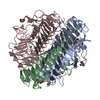
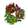
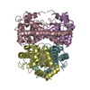
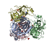
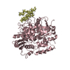
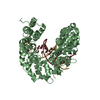
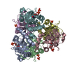
| Title | Crystal structure of Serine acetyltransferase isoform 3 in complex with cysteine from Entamoeba histolytica | ||||||
 Components Components | Serine acetyltransferase 1, putative | ||||||
 Keywords Keywords | TRANSFERASE / Left handed beta-helical (LBH) domain / serine acetyltransferase / Cysteine biosynthesis / Cysteine synthase Complex. | ||||||
| Function / homology |  Function and homology information Function and homology informationserine O-acetyltransferase / serine O-acetyltransferase activity / amino acid biosynthetic process Similarity search - Function | ||||||
| Biological species |   Entamoeba histolytica (eukaryote) Entamoeba histolytica (eukaryote) | ||||||
| Method |  X-RAY DIFFRACTION / SYNCHROTRON / MOLECULAR REPLACEMENT / Resolution: 2.131 Å X-RAY DIFFRACTION / SYNCHROTRON / MOLECULAR REPLACEMENT / Resolution: 2.131 Å | ||||||
 Authors Authors | Dharavath, S. / Kumar, S. / Gourinath, S. | ||||||
| Funding support |  India, 1items India, 1items
| ||||||
 Citation Citation | Journal: To Be Published Title: Crystal structure of Serine acetyltransferase isoform 3 in complex with cysteine from Entamoeba histolytica Authors: Dharavath, S. / Kumar, S. | ||||||
| History |
|
- Structure visualization
| Structure viewer | Molecule: MolmilJmol/JSmol |
|---|
- Downloads & links
-Download
| PDBx/mmCIF format | 7bw9.cif.gz | 181.9 KB | Display | PDBx/mmCIF format |
|---|---|---|---|---|
| PDB format | pdb7bw9.ent.gz | 142.4 KB | Display | PDB format |
| PDBx/mmJSON format | 7bw9.json.gz | Tree view | PDBx/mmJSON format | |
| Others |  Other downloads Other downloads |
-Validation report
| Summary document | 7bw9_validation.pdf.gz | 1.3 MB | Display | wwPDB validaton report |
|---|---|---|---|---|
| Full document | 7bw9_full_validation.pdf.gz | 1.3 MB | Display | |
| Data in XML | 7bw9_validation.xml.gz | 34 KB | Display | |
| Data in CIF | 7bw9_validation.cif.gz | 48.5 KB | Display | |
| Arichive directory | https://data.pdbj.org/pub/pdb/validation_reports/bw/7bw9ftp://data.pdbj.org/pub/pdb/validation_reports/bw/7bw9 | HTTPS FTP |
-Related structure data
| Related structure data |  3p47S S: Starting model for refinement |
|---|---|
| Similar structure data |
-Links
 PDBj
PDBj


- Assembly
| Deposited unit | 
| ||||||||
|---|---|---|---|---|---|---|---|---|---|
| 1 |
| ||||||||
| Unit cell |
| ||||||||
| Components on special symmetry positions |
|
-Components
| #1: Protein | Mass: 37738.914 Da / Num. of mol.: 3 Source method: isolated from a genetically manipulated source Source: (gene. exp.) Entamoeba histolytica (eukaryote) / Gene: KM1_023830 / Production host:  #2: Chemical |   Type: L-peptide linking / Mass: 121.158 Da / Num. of mol.: 3 / Source method: obtained synthetically / Formula: C3H7NO2S / Feature type: SUBJECT OF INVESTIGATION Type: L-peptide linking / Mass: 121.158 Da / Num. of mol.: 3 / Source method: obtained synthetically / Formula: C3H7NO2S / Feature type: SUBJECT OF INVESTIGATION#3: Water | ChemComp-HOH / |  Mass: 18.015 Da / Num. of mol.: 356 / Source method: isolated from a natural source / Formula: H2O Mass: 18.015 Da / Num. of mol.: 356 / Source method: isolated from a natural source / Formula: H2OHas ligand of interest | Y | |
|---|
-Experimental details
-Experiment
| Experiment | Method: X-RAY DIFFRACTION / Number of used crystals: 1 |
|---|
- Sample preparation
| Crystal | Density Matthews: 1.99 Å3/Da / Density % sol: 38.2 % |
|---|---|
| Crystal grow | Temperature: 289.15 K / Method: vapor diffusion, hanging drop / pH: 7.5 Details: 0.1 M Hepes pH 7.5, 0.2 M KSCN, 0.2 M KBr, 25 % PEG 3350 PH range: 7.2 - 8.5 |
-Data collection
| Diffraction | Mean temperature: 193 K / Serial crystal experiment: N |
|---|---|
| Diffraction source | Source: SYNCHROTRON / Site: ESRF  / Beamline: BM14 / Wavelength: 1 Å / Beamline: BM14 / Wavelength: 1 Å |
| Detector | Type: MARMOSAIC 225 mm CCD / Detector: CCD / Date: Apr 28, 2014 |
| Radiation | Monochromator: Si(111) monochromator / Protocol: SINGLE WAVELENGTH / Monochromatic (M) / Laue (L): M / Scattering type: x-ray |
| Radiation wavelength | Wavelength: 1 Å / Relative weight: 1 |
| Reflection | Resolution: 2.13→50 Å / Num. obs: 48708 / % possible obs: 99.9 % / Redundancy: 5.2 % / CC1/2: 0.95 / CC star: 0.98 / Rpim(I) all: 0.053 / Χ2: 1.054 / Net I/σ(I): 15.3 |
| Reflection shell | Resolution: 2.13→2.17 Å / Redundancy: 4.6 % / Mean I/σ(I) obs: 2.2 / Num. unique obs: 2424 / CC1/2: 0.82 / CC star: 0.95 / Rpim(I) all: 0.28 / Χ2: 0.767 / % possible all: 99.5 |
- Processing
| Software |
| ||||||||||||||||||||||||||||||||||||||||||||||||
|---|---|---|---|---|---|---|---|---|---|---|---|---|---|---|---|---|---|---|---|---|---|---|---|---|---|---|---|---|---|---|---|---|---|---|---|---|---|---|---|---|---|---|---|---|---|---|---|---|---|
| Refinement | Method to determine structure: MOLECULAR REPLACEMENT Starting model: 3p47 Resolution: 2.131→39.54 Å / SU ML: 0.26 / Cross valid method: THROUGHOUT / σ(F): 1.38 / Phase error: 23.26 / Stereochemistry target values: ML
| ||||||||||||||||||||||||||||||||||||||||||||||||
| Solvent computation | Shrinkage radii: 0.9 Å / VDW probe radii: 1.11 Å / Solvent model: FLAT BULK SOLVENT MODEL | ||||||||||||||||||||||||||||||||||||||||||||||||
| Displacement parameters | Biso max: 84.28 Å2 / Biso mean: 27.8204 Å2 / Biso min: 8.12 Å2 | ||||||||||||||||||||||||||||||||||||||||||||||||
| Refinement step | Cycle: final / Resolution: 2.131→39.54 Å
| ||||||||||||||||||||||||||||||||||||||||||||||||
| LS refinement shell | Refine-ID: X-RAY DIFFRACTION / Rfactor Rfree error: 0
|
