 Movie
Movie Controller

[English] 日本語
 Yorodumi
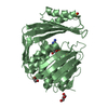
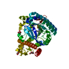
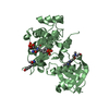
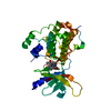
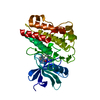
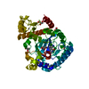
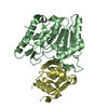
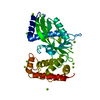
- PDB-7bvk: UDP-N-acetylglucosamine 3-dehydrogenase GnnA from Acidithiobacill... -
+ Open data

- Basic information
| Entry | Database: PDB / ID: 7bvk | ||||||
|---|---|---|---|---|---|---|---|
| Title | UDP-N-acetylglucosamine 3-dehydrogenase GnnA from Acidithiobacillus ferrooxidans (P212121) | ||||||
 Components Components | Oxidoreductase, NAD-binding | ||||||
 Keywords Keywords | OXIDOREDUCTASE / GnnA / UDP-GlcNAc / lipopolysaccharide / UDP-N-acetylglucosamine 3-dehydrogenase | ||||||
| Function / homology |  Function and homology information Function and homology informationUDP-N-acetylglucosamine 3-dehydrogenase / lipid A biosynthetic process / oxidoreductase activity / nucleotide binding / membrane Similarity search - Function | ||||||
| Biological species |  Acidithiobacillus ferrooxidans (bacteria) Acidithiobacillus ferrooxidans (bacteria) | ||||||
| Method |  X-RAY DIFFRACTION / SYNCHROTRON / MOLECULAR REPLACEMENT / molecular replacement / Resolution: 2.7 Å X-RAY DIFFRACTION / SYNCHROTRON / MOLECULAR REPLACEMENT / molecular replacement / Resolution: 2.7 Å | ||||||
 Authors Authors | Wangkanont, K. | ||||||
| Funding support |  Thailand, 1items Thailand, 1items
| ||||||
 Citation Citation | Journal: Acs Chem.Biol. / Year: 2020 Title: Biochemical and Structural Investigation of GnnA in the Lipopolysaccharide Biosynthesis Pathway of Acidithiobacillus ferrooxidans . Authors: Manissorn, J. / Sitthiyotha, T. / Montalban, J.R.E. / Chunsrivirot, S. / Thongnuek, P. / Wangkanont, K. | ||||||
| History |
|
- Structure visualization
| Structure viewer | Molecule: MolmilJmol/JSmol |
|---|
- Downloads & links
-Download
| PDBx/mmCIF format | 7bvk.cif.gz | 131.1 KB | Display | PDBx/mmCIF format |
|---|---|---|---|---|
| PDB format | pdb7bvk.ent.gz | 100.2 KB | Display | PDB format |
| PDBx/mmJSON format | 7bvk.json.gz | Tree view | PDBx/mmJSON format | |
| Others |  Other downloads Other downloads |
-Validation report
| Arichive directory | https://data.pdbj.org/pub/pdb/validation_reports/bv/7bvkftp://data.pdbj.org/pub/pdb/validation_reports/bv/7bvk | HTTPS FTP |
|---|
-Related structure data
| Related structure data |  7bvjSC S: Starting model for refinement C: citing same article ( |
|---|---|
| Similar structure data |

-Links
 PDBj
PDBj- Assembly
| Deposited unit | 
| ||||||||
|---|---|---|---|---|---|---|---|---|---|
| 1 | 
| ||||||||
| 2 | 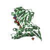
| ||||||||
| Unit cell |
|
-Components
| #1: Protein | Mass: 35163.395 Da / Num. of mol.: 2 Source method: isolated from a genetically manipulated source Details: synthetic gene, codon optimized Source: (gene. exp.) Acidithiobacillus ferrooxidans (strain ATCC 23270 / DSM 14882 / CIP 104768 / NCIMB 8455) (bacteria)Strain: ATCC 23270 / DSM 14882 / CIP 104768 / NCIMB 8455 / Gene: AFE_1457 / Plasmid: pET24a / Production host: #2: Chemical |   Mass: 663.425 Da / Num. of mol.: 2 / Source method: obtained synthetically / Formula: C21H27N7O14P2 / Feature type: SUBJECT OF INVESTIGATION / Comment: NAD*YM Mass: 663.425 Da / Num. of mol.: 2 / Source method: obtained synthetically / Formula: C21H27N7O14P2 / Feature type: SUBJECT OF INVESTIGATION / Comment: NAD*YM#3: Chemical |   Mass: 96.063 Da / Num. of mol.: 3 / Source method: obtained synthetically / Formula: SO4 Mass: 96.063 Da / Num. of mol.: 3 / Source method: obtained synthetically / Formula: SO4#4: Water | ChemComp-HOH / |  Mass: 18.015 Da / Num. of mol.: 64 / Source method: isolated from a natural source / Formula: H2O Mass: 18.015 Da / Num. of mol.: 64 / Source method: isolated from a natural source / Formula: H2OHas ligand of interest | Y | |
|---|
-Experimental details
-Experiment
| Experiment | Method: X-RAY DIFFRACTION / Number of used crystals: 1 |
|---|
- Sample preparation
| Crystal | Density Matthews: 2.19 Å3/Da / Density % sol: 43.94 % / Mosaicity: 0.28 ° |
|---|---|
| Crystal grow | Temperature: 289 K / Method: batch mode / pH: 5.5 Details: 100 mM Bis-Tris, 200 mM Ammonium Sulfate, 25% PEG 3350 |
-Data collection
| Diffraction | Mean temperature: 100 K / Serial crystal experiment: N | ||||||||||||||||||||||||||||||
|---|---|---|---|---|---|---|---|---|---|---|---|---|---|---|---|---|---|---|---|---|---|---|---|---|---|---|---|---|---|---|---|
| Diffraction source | Source: SYNCHROTRON / Site: NSRRC  / Beamline: BL13B1 / Wavelength: 0.999999 Å / Beamline: BL13B1 / Wavelength: 0.999999 Å | ||||||||||||||||||||||||||||||
| Detector | Type: ADSC QUANTUM 315r / Detector: CCD / Date: Dec 20, 2019 | ||||||||||||||||||||||||||||||
| Radiation | Monochromator: Si(111) / Protocol: SINGLE WAVELENGTH / Monochromatic (M) / Laue (L): M / Scattering type: x-ray | ||||||||||||||||||||||||||||||
| Radiation wavelength | Wavelength: 0.999999 Å / Relative weight: 1 | ||||||||||||||||||||||||||||||
| Reflection | Resolution: 2.7→19.76 Å / Num. obs: 17552 / % possible obs: 99.7 % / Redundancy: 7.1 % / CC1/2: 0.99 / Rmerge(I) obs: 0.213 / Rpim(I) all: 0.086 / Rrim(I) all: 0.23 / Net I/σ(I): 6.5 / Num. measured all: 125291 / Scaling rejects: 86 | ||||||||||||||||||||||||||||||
| Reflection shell | Diffraction-ID: 1
|
-Phasing
| Phasing | Method: molecular replacement | |||||||||
|---|---|---|---|---|---|---|---|---|---|---|
| Phasing MR |
|
- Processing
| Software |
| ||||||||||||||||||||||||||||||||||||||||||||||||||||||||
|---|---|---|---|---|---|---|---|---|---|---|---|---|---|---|---|---|---|---|---|---|---|---|---|---|---|---|---|---|---|---|---|---|---|---|---|---|---|---|---|---|---|---|---|---|---|---|---|---|---|---|---|---|---|---|---|---|---|
| Refinement | Method to determine structure: MOLECULAR REPLACEMENT Starting model: 7BVJ Resolution: 2.7→19.76 Å / SU ML: 0.4 / Cross valid method: THROUGHOUT / σ(F): 1.36 / Phase error: 34.63 / Stereochemistry target values: ML
| ||||||||||||||||||||||||||||||||||||||||||||||||||||||||
| Solvent computation | Shrinkage radii: 0.9 Å / VDW probe radii: 1.11 Å / Solvent model: FLAT BULK SOLVENT MODEL | ||||||||||||||||||||||||||||||||||||||||||||||||||||||||
| Displacement parameters | Biso max: 115.14 Å2 / Biso mean: 42.2284 Å2 / Biso min: 16.83 Å2 | ||||||||||||||||||||||||||||||||||||||||||||||||||||||||
| Refinement step | Cycle: final / Resolution: 2.7→19.76 Å
| ||||||||||||||||||||||||||||||||||||||||||||||||||||||||
| LS refinement shell | Refine-ID: X-RAY DIFFRACTION / Rfactor Rfree error: 0 / Total num. of bins used: 7
|
