 Movie
Movie Controller

[English] 日本語
 Yorodumi
- PDB-7bva: Crystal structure of UDP-N-acetylmuramic Acid L-alanine ligase (M... -
+ Open data

- Basic information
| Entry | Database: PDB / ID: 7bva | ||||||
|---|---|---|---|---|---|---|---|
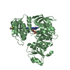
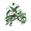
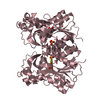
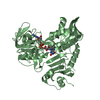
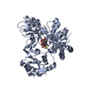
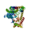
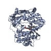
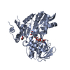
| Title | Crystal structure of UDP-N-acetylmuramic Acid L-alanine ligase (MurC) from Mycobacterium bovis | ||||||
 Components Components | UDP-N-acetylmuramate--L-alanine ligase | ||||||
 Keywords Keywords | LIGASE / MurC / Peptidoglycan | ||||||
| Function / homology |  Function and homology information Function and homology informationUDP-N-acetylmuramate-L-alanine ligase / UDP-N-acetylmuramate-L-alanine ligase activity / peptidoglycan biosynthetic process / cell wall organization / regulation of cell shape / cell division / ATP binding / cytoplasm Similarity search - Function | ||||||
| Biological species |  Mycobacterium bovis (bacteria) Mycobacterium bovis (bacteria) | ||||||
| Method |  X-RAY DIFFRACTION / SYNCHROTRON / MOLECULAR REPLACEMENT / Resolution: 2.303 Å X-RAY DIFFRACTION / SYNCHROTRON / MOLECULAR REPLACEMENT / Resolution: 2.303 Å | ||||||
 Authors Authors | Seo, P.W. / Kim, J.S. | ||||||
| Funding support |  Korea, Republic Of, 1items Korea, Republic Of, 1items
| ||||||
 Citation Citation | Journal: Acta Crystallogr D Struct Biol / Year: 2021 Title: Crystal structures of UDP-N-acetylmuramic acid L-alanine ligase (MurC) from Mycobacterium bovis with and without UDP-N-acetylglucosamine. Authors: Seo, P.W. / Park, S.Y. / Hofmann, A. / Kim, J.S. | ||||||
| History |
|
- Structure visualization
| Structure viewer | Molecule: MolmilJmol/JSmol |
|---|
- Downloads & links
-Download
| PDBx/mmCIF format | 7bva.cif.gz | 193.2 KB | Display | PDBx/mmCIF format |
|---|---|---|---|---|
| PDB format | pdb7bva.ent.gz | 149.3 KB | Display | PDB format |
| PDBx/mmJSON format | 7bva.json.gz | Tree view | PDBx/mmJSON format | |
| Others |  Other downloads Other downloads |
-Validation report
| Arichive directory | https://data.pdbj.org/pub/pdb/validation_reports/bv/7bvaftp://data.pdbj.org/pub/pdb/validation_reports/bv/7bva | HTTPS FTP |
|---|
-Related structure data
| Related structure data | 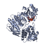 7bvbC 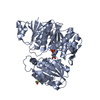 4hv4S S: Starting model for refinement C: citing same article ( |
|---|---|
| Similar structure data |


-Links
 PDBj
PDBj- Assembly
| Deposited unit | 
| ||||||||
|---|---|---|---|---|---|---|---|---|---|
| 1 | 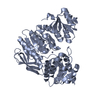
| ||||||||
| 2 | 
| ||||||||
| Unit cell |
|
-Components
| #1: Protein | Mass: 54402.746 Da / Num. of mol.: 2 Source method: isolated from a genetically manipulated source Source: (gene. exp.) Mycobacterium bovis (strain ATCC BAA-935 / AF2122/97) (bacteria)Gene: murC, BQ2027_MB2176C / Production host: References: UniProt: P65473, UDP-N-acetylmuramate-L-alanine ligase #2: Chemical |   Mass: 65.409 Da / Num. of mol.: 2 / Source method: obtained synthetically / Formula: Zn / Feature type: SUBJECT OF INVESTIGATION Mass: 65.409 Da / Num. of mol.: 2 / Source method: obtained synthetically / Formula: Zn / Feature type: SUBJECT OF INVESTIGATION#3: Water | ChemComp-HOH / |  Mass: 18.015 Da / Num. of mol.: 301 / Source method: isolated from a natural source / Formula: H2O Mass: 18.015 Da / Num. of mol.: 301 / Source method: isolated from a natural source / Formula: H2OHas ligand of interest | Y | |
|---|
-Experimental details
-Experiment
| Experiment | Method: X-RAY DIFFRACTION / Number of used crystals: 1 |
|---|
- Sample preparation
| Crystal | Density Matthews: 2.3 Å3/Da / Density % sol: 46.53 % |
|---|---|
| Crystal grow | Temperature: 294 K / Method: vapor diffusion, hanging drop Details: 0.1M HEPES pH 7.5, 0.2M NaCl, 30% (w/v) PEG 1500, 5% (v/v) MPD |
-Data collection
| Diffraction | Mean temperature: 100 K / Serial crystal experiment: N |
|---|---|
| Diffraction source | Source: SYNCHROTRON / Site: PAL/PLS / Beamline: 5C (4A) / Wavelength: 0.9796 Å |
| Detector | Type: ADSC QUANTUM 315r / Detector: CCD / Date: Sep 19, 2017 |
| Radiation | Protocol: SINGLE WAVELENGTH / Monochromatic (M) / Laue (L): M / Scattering type: x-ray |
| Radiation wavelength | Wavelength: 0.9796 Å / Relative weight: 1 |
| Reflection | Resolution: 2.3→50 Å / Num. obs: 43282 / % possible obs: 99 % / Redundancy: 2.6 % / Biso Wilson estimate: 32.42 Å2 / Rsym value: 0.087 / Net I/σ(I): 10.9 |
| Reflection shell | Resolution: 2.3→2.34 Å / Num. unique obs: 2172 / Rsym value: 0.607 |
- Processing
| Software |
| ||||||||||||||||||||||||||||||||||||||||||||||||||||||||||||||||||||||||||||||||||||||||||||||||||||||||||||||||||||||||
|---|---|---|---|---|---|---|---|---|---|---|---|---|---|---|---|---|---|---|---|---|---|---|---|---|---|---|---|---|---|---|---|---|---|---|---|---|---|---|---|---|---|---|---|---|---|---|---|---|---|---|---|---|---|---|---|---|---|---|---|---|---|---|---|---|---|---|---|---|---|---|---|---|---|---|---|---|---|---|---|---|---|---|---|---|---|---|---|---|---|---|---|---|---|---|---|---|---|---|---|---|---|---|---|---|---|---|---|---|---|---|---|---|---|---|---|---|---|---|---|---|---|
| Refinement | Method to determine structure: MOLECULAR REPLACEMENT Starting model: 4HV4 Resolution: 2.303→32.727 Å / SU ML: 0.27 / Cross valid method: THROUGHOUT / σ(F): 1.34 / Phase error: 24.04
| ||||||||||||||||||||||||||||||||||||||||||||||||||||||||||||||||||||||||||||||||||||||||||||||||||||||||||||||||||||||||
| Solvent computation | Shrinkage radii: 0.9 Å / VDW probe radii: 1.11 Å | ||||||||||||||||||||||||||||||||||||||||||||||||||||||||||||||||||||||||||||||||||||||||||||||||||||||||||||||||||||||||
| Displacement parameters | Biso max: 121.29 Å2 / Biso mean: 40.1981 Å2 / Biso min: 15.49 Å2 | ||||||||||||||||||||||||||||||||||||||||||||||||||||||||||||||||||||||||||||||||||||||||||||||||||||||||||||||||||||||||
| Refinement step | Cycle: final / Resolution: 2.303→32.727 Å
| ||||||||||||||||||||||||||||||||||||||||||||||||||||||||||||||||||||||||||||||||||||||||||||||||||||||||||||||||||||||||
| Refine LS restraints |
| ||||||||||||||||||||||||||||||||||||||||||||||||||||||||||||||||||||||||||||||||||||||||||||||||||||||||||||||||||||||||
| LS refinement shell | Refine-ID: X-RAY DIFFRACTION / Rfactor Rfree error: 0
|
