 Movie
Movie Controller

[English] 日本語
 Yorodumi
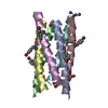
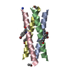
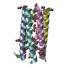
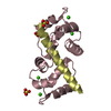
- PDB-7bo8: A hexameric de novo coiled-coil assembly: CC-Type2-(VaYd)4-Y3F-W1... -
+ Open data

- Basic information
| Entry | Database: PDB / ID: 7bo8 | |||||||||
|---|---|---|---|---|---|---|---|---|---|---|
| Title | A hexameric de novo coiled-coil assembly: CC-Type2-(VaYd)4-Y3F-W19(BrPhe)-Y24F. | |||||||||
 Components Components | CC-Type2-(VaYd)4-Y3F-W19(BrPhe)-Y24F | |||||||||
 Keywords Keywords | DE NOVO PROTEIN / Coiled coil / synthetic peptide / homomeric assembly / tyrosine-tyrosine interactions | |||||||||
| Function / homology | OXAMIC ACID Function and homology information Function and homology information | |||||||||
| Biological species | synthetic construct (others) | |||||||||
| Method |  X-RAY DIFFRACTION / SYNCHROTRON / SAD / molecular replacement / Resolution: 1.84 Å X-RAY DIFFRACTION / SYNCHROTRON / SAD / molecular replacement / Resolution: 1.84 Å | |||||||||
 Authors Authors | Rhys, G.G. / Brady, R.L. / Woolfson, D.N. | |||||||||
| Funding support | European Union,  United Kingdom, 2items United Kingdom, 2items
| |||||||||
 Citation Citation | Journal: Biomacromolecules / Year: 2021 Title: How Coiled-Coil Assemblies Accommodate Multiple Aromatic Residues. Authors: Rhys, G.G. / Dawson, W.M. / Beesley, J.L. / Martin, F.J.O. / Brady, R.L. / Thomson, A.R. / Woolfson, D.N. | |||||||||
| History |
|
- Structure visualization
| Structure viewer | Molecule: MolmilJmol/JSmol |
|---|
- Downloads & links
-Download
| PDBx/mmCIF format | 7bo8.cif.gz | 46.3 KB | Display | PDBx/mmCIF format |
|---|---|---|---|---|
| PDB format | pdb7bo8.ent.gz | 35.1 KB | Display | PDB format |
| PDBx/mmJSON format | 7bo8.json.gz | Tree view | PDBx/mmJSON format | |
| Others |  Other downloads Other downloads |
-Validation report
| Summary document | 7bo8_validation.pdf.gz | 487.3 KB | Display | wwPDB validaton report |
|---|---|---|---|---|
| Full document | 7bo8_full_validation.pdf.gz | 487.5 KB | Display | |
| Data in XML | 7bo8_validation.xml.gz | 8.8 KB | Display | |
| Data in CIF | 7bo8_validation.cif.gz | 11.7 KB | Display | |
| Arichive directory | https://data.pdbj.org/pub/pdb/validation_reports/bo/7bo8ftp://data.pdbj.org/pub/pdb/validation_reports/bo/7bo8 | HTTPS FTP |
-Related structure data


-Links
 PDBj
PDBj

- Assembly
| Deposited unit | 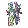
| ||||||||
|---|---|---|---|---|---|---|---|---|---|
| 1 |
| ||||||||
| Unit cell |
|
-Components
| #1: Protein/peptide | Mass: 3386.687 Da / Num. of mol.: 6 / Source method: obtained synthetically / Source: (synth.) synthetic construct (others) #2: Chemical | ChemComp-OXM / |   Mass: 89.050 Da / Num. of mol.: 1 / Source method: obtained synthetically / Formula: C2H3NO3 Mass: 89.050 Da / Num. of mol.: 1 / Source method: obtained synthetically / Formula: C2H3NO3#3: Chemical | ChemComp-EDO / |   Mass: 62.068 Da / Num. of mol.: 1 / Source method: obtained synthetically / Formula: C2H6O2 Mass: 62.068 Da / Num. of mol.: 1 / Source method: obtained synthetically / Formula: C2H6O2#4: Water | ChemComp-HOH / |  Mass: 18.015 Da / Num. of mol.: 32 / Source method: isolated from a natural source / Formula: H2O Mass: 18.015 Da / Num. of mol.: 32 / Source method: isolated from a natural source / Formula: H2OHas ligand of interest | N | Has protein modification | Y | |
|---|
-Experimental details
-Experiment
| Experiment | Method: X-RAY DIFFRACTION / Number of used crystals: 1 |
|---|
- Sample preparation
| Crystal | Density Matthews: 2.28 Å3/Da / Density % sol: 46.13 % |
|---|---|
| Crystal grow | Temperature: 292 K / Method: vapor diffusion, sitting drop / pH: 8.5 Details: After 1:1 dilution with the peptide solution the resulting conditions were 0.05 M Sodium formate, 0.05 M Ammonium acetate, 0.05 M Sodium citrate tribasic dihydrate, 0.05 M Potassium sodium ...Details: After 1:1 dilution with the peptide solution the resulting conditions were 0.05 M Sodium formate, 0.05 M Ammonium acetate, 0.05 M Sodium citrate tribasic dihydrate, 0.05 M Potassium sodium tartrate tetrahydrate, 0.05 M Sodium oxamate, 0.05 M Tris, 0.05 M BICINE, 6% v/v Ethylene glycol and 3% w/v PEG 8000 |
-Data collection
| Diffraction | Mean temperature: 80 K / Serial crystal experiment: N | ||||||||||||||||||||||||||||||
|---|---|---|---|---|---|---|---|---|---|---|---|---|---|---|---|---|---|---|---|---|---|---|---|---|---|---|---|---|---|---|---|
| Diffraction source | Source: SYNCHROTRON / Site: Diamond / Beamline: I03 / Wavelength: 0.91983 Å | ||||||||||||||||||||||||||||||
| Detector | Type: DECTRIS PILATUS3 6M / Detector: PIXEL / Date: Oct 5, 2017 | ||||||||||||||||||||||||||||||
| Radiation | Protocol: SINGLE WAVELENGTH / Monochromatic (M) / Laue (L): M / Scattering type: x-ray | ||||||||||||||||||||||||||||||
| Radiation wavelength | Wavelength: 0.91983 Å / Relative weight: 1 | ||||||||||||||||||||||||||||||
| Reflection | Resolution: 1.84→38.57 Å / Num. obs: 16884 / % possible obs: 99.9 % / Redundancy: 32.1 % / Biso Wilson estimate: 45.32 Å2 / CC1/2: 0.998 / Rmerge(I) obs: 0.176 / Rpim(I) all: 0.03 / Rrim(I) all: 0.176 / Net I/σ(I): 11.3 | ||||||||||||||||||||||||||||||
| Reflection shell | Diffraction-ID: 1
|
-Phasing
| Phasing | Method: molecular replacement |
|---|
- Processing
| Software |
| |||||||||||||||||||||||||||||||||||||||||||||||||||||||
|---|---|---|---|---|---|---|---|---|---|---|---|---|---|---|---|---|---|---|---|---|---|---|---|---|---|---|---|---|---|---|---|---|---|---|---|---|---|---|---|---|---|---|---|---|---|---|---|---|---|---|---|---|---|---|---|---|
| Refinement | Method to determine structure: SAD / Resolution: 1.84→38.57 Å / Cor.coef. Fo:Fc: 0.967 / Cor.coef. Fo:Fc free: 0.943 / SU B: 4.208 / SU ML: 0.12 / Cross valid method: THROUGHOUT / σ(F): 0 / ESU R: 0.143 / ESU R Free: 0.151 / Stereochemistry target values: MAXIMUM LIKELIHOOD Details: HYDROGENS HAVE BEEN ADDED IN THE RIDING POSITIONS U VALUES : REFINED INDIVIDUALLY
| |||||||||||||||||||||||||||||||||||||||||||||||||||||||
| Solvent computation | Ion probe radii: 0.8 Å / Shrinkage radii: 0.8 Å / VDW probe radii: 1.2 Å / Solvent model: MASK | |||||||||||||||||||||||||||||||||||||||||||||||||||||||
| Displacement parameters | Biso max: 107.15 Å2 / Biso mean: 47.797 Å2 / Biso min: 32.22 Å2
| |||||||||||||||||||||||||||||||||||||||||||||||||||||||
| Refinement step | Cycle: final / Resolution: 1.84→38.57 Å
| |||||||||||||||||||||||||||||||||||||||||||||||||||||||
| Refine LS restraints |
| |||||||||||||||||||||||||||||||||||||||||||||||||||||||
| LS refinement shell | Resolution: 1.84→1.888 Å / Rfactor Rfree error: 0 / Total num. of bins used: 20
|
