 Movie
Movie Controller

[English] 日本語
 Yorodumi
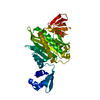
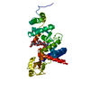
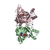
- PDB-7bkf: Crystal structure of WT BA3943, a CE4 family pseudoenzyme from Ba... -
+ Open data

- Basic information
| Entry | Database: PDB / ID: 7bkf | ||||||
|---|---|---|---|---|---|---|---|
| Title | Crystal structure of WT BA3943, a CE4 family pseudoenzyme from Bacillus Anthracis | ||||||
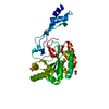
 Components Components | Putative polysaccharide deacetylase | ||||||
 Keywords Keywords | UNKNOWN FUNCTION / Polysaccharide / Deacetylase / Peptidoglycan / Pseudoenzyme / Sporulation. | ||||||
| Function / homology | Sporulation protein, polysaccharide deacetylase, YlxY / NodB homology domain profile. / NodB homology domain / Polysaccharide deacetylase / hydrolase activity, acting on carbon-nitrogen (but not peptide) bonds / Glycoside hydrolase/deacetylase, beta/alpha-barrel / carbohydrate metabolic process / Putative polysaccharide deacetylase Function and homology information Function and homology information | ||||||
| Biological species |  | ||||||
| Method |  X-RAY DIFFRACTION / SYNCHROTRON / MOLECULAR REPLACEMENT / Resolution: 1.139 Å X-RAY DIFFRACTION / SYNCHROTRON / MOLECULAR REPLACEMENT / Resolution: 1.139 Å | ||||||
 Authors Authors | Molfetas, A. / Kokkinidis, M. | ||||||
| Funding support |  Greece, 1items Greece, 1items
| ||||||
 Citation Citation | Journal: To Be Published Title: The resurrection of a dead enzyme Authors: Molfetas, A. / Tomatsidou, N. / Bouriotis, V. / Kokkinidis, M. | ||||||
| History |
|
- Structure visualization
| Structure viewer | Molecule: MolmilJmol/JSmol |
|---|
- Downloads & links
-Download
| PDBx/mmCIF format | 7bkf.cif.gz | 142.2 KB | Display | PDBx/mmCIF format |
|---|---|---|---|---|
| PDB format | pdb7bkf.ent.gz | 110.6 KB | Display | PDB format |
| PDBx/mmJSON format | 7bkf.json.gz | Tree view | PDBx/mmJSON format | |
| Others |  Other downloads Other downloads |
-Validation report
| Arichive directory | https://data.pdbj.org/pub/pdb/validation_reports/bk/7bkfftp://data.pdbj.org/pub/pdb/validation_reports/bk/7bkf | HTTPS FTP |
|---|
-Related structure data
| Related structure data |  6hpaS S: Starting model for refinement |
|---|---|
| Similar structure data |


-Links
 PDBj
PDBj
- Assembly
| Deposited unit | 
| ||||||||
|---|---|---|---|---|---|---|---|---|---|
| 1 |
| ||||||||
| Unit cell |
|
-Components
| #1: Protein | Mass: 32910.676 Da / Num. of mol.: 1 Source method: isolated from a genetically manipulated source Source: (gene. exp.) | ||||||
|---|---|---|---|---|---|---|---|
| #2: Chemical | ChemComp-SO4 /   Mass: 96.063 Da / Num. of mol.: 4 / Source method: obtained synthetically / Formula: SO4 Mass: 96.063 Da / Num. of mol.: 4 / Source method: obtained synthetically / Formula: SO4#3: Chemical | ChemComp-TRS / |   Mass: 122.143 Da / Num. of mol.: 1 / Source method: obtained synthetically / Formula: C4H12NO3 / Comment: pH buffer*YM Mass: 122.143 Da / Num. of mol.: 1 / Source method: obtained synthetically / Formula: C4H12NO3 / Comment: pH buffer*YM#4: Water | ChemComp-HOH / |  Mass: 18.015 Da / Num. of mol.: 249 / Source method: isolated from a natural source / Formula: H2O Mass: 18.015 Da / Num. of mol.: 249 / Source method: isolated from a natural source / Formula: H2OHas ligand of interest | N | |
-Experimental details
-Experiment
| Experiment | Method: X-RAY DIFFRACTION / Number of used crystals: 1 |
|---|
- Sample preparation
| Crystal | Density Matthews: 2.09 Å3/Da / Density % sol: 41.06 % |
|---|---|
| Crystal grow | Temperature: 293 K / Method: vapor diffusion, hanging drop / pH: 5 / Details: (NH4)2SO4 CH3COONA PH=5 / PH range: 5.0-6.0 |
-Data collection
| Diffraction | Mean temperature: 100 K / Serial crystal experiment: N |
|---|---|
| Diffraction source | Source: SYNCHROTRON / Site: BESSY  / Beamline: 14.2 / Wavelength: 0.9184 Å / Beamline: 14.2 / Wavelength: 0.9184 Å |
| Detector | Type: DECTRIS PILATUS 2M / Detector: PIXEL / Date: Apr 6, 2017 |
| Radiation | Monochromator: DCM Si(111) / Protocol: SINGLE WAVELENGTH / Monochromatic (M) / Laue (L): M / Scattering type: x-ray |
| Radiation wavelength | Wavelength: 0.9184 Å / Relative weight: 1 |
| Reflection | Resolution: 1.139→40.7 Å / Num. obs: 92525 / % possible obs: 96.1 % / Redundancy: 6.0872 % / Biso Wilson estimate: 19.3 Å2 / Rrim(I) all: 0.075 / Net I/σ(I): 11.14 |
| Reflection shell | Resolution: 1.14→1.21 Å / Mean I/σ(I) obs: 0.33 / Num. unique obs: 12163 / CC1/2: 0.16 / % possible all: 79 |
- Processing
| Software |
| ||||||||||||||||||||||||||||||||||||||||||||||||||||||||||||||||||||||||||||||||||||||||||||||||
|---|---|---|---|---|---|---|---|---|---|---|---|---|---|---|---|---|---|---|---|---|---|---|---|---|---|---|---|---|---|---|---|---|---|---|---|---|---|---|---|---|---|---|---|---|---|---|---|---|---|---|---|---|---|---|---|---|---|---|---|---|---|---|---|---|---|---|---|---|---|---|---|---|---|---|---|---|---|---|---|---|---|---|---|---|---|---|---|---|---|---|---|---|---|---|---|---|---|
| Refinement | Method to determine structure: MOLECULAR REPLACEMENT Starting model: 6HPA Resolution: 1.139→40.656 Å / SU ML: 0.2 / Cross valid method: THROUGHOUT / σ(F): 1.32 / Phase error: 27.37 / Stereochemistry target values: ML
| ||||||||||||||||||||||||||||||||||||||||||||||||||||||||||||||||||||||||||||||||||||||||||||||||
| Solvent computation | Shrinkage radii: 0.9 Å / VDW probe radii: 1.11 Å / Solvent model: FLAT BULK SOLVENT MODEL | ||||||||||||||||||||||||||||||||||||||||||||||||||||||||||||||||||||||||||||||||||||||||||||||||
| Displacement parameters | Biso max: 77.78 Å2 / Biso mean: 24.2257 Å2 / Biso min: 12.28 Å2 | ||||||||||||||||||||||||||||||||||||||||||||||||||||||||||||||||||||||||||||||||||||||||||||||||
| Refinement step | Cycle: final / Resolution: 1.139→40.656 Å
| ||||||||||||||||||||||||||||||||||||||||||||||||||||||||||||||||||||||||||||||||||||||||||||||||
| Refine LS restraints |
| ||||||||||||||||||||||||||||||||||||||||||||||||||||||||||||||||||||||||||||||||||||||||||||||||
| LS refinement shell | Refine-ID: X-RAY DIFFRACTION / Rfactor Rfree error: 0
|
