 Movie
Movie Controller

[English] 日本語
 Yorodumi
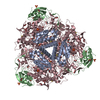
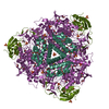
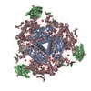
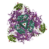
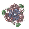
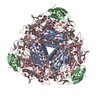
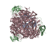
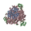
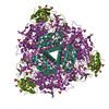
- PDB-7b5a: X-ray crystal structure of Sporosarcina pasteurii urease inhibite... -
+ Open data

- Basic information
| Entry | Database: PDB / ID: 7b5a | ||||||
|---|---|---|---|---|---|---|---|
| Title | X-ray crystal structure of Sporosarcina pasteurii urease inhibited by Ag(PEt3)2NO3 determined at 1.97 Angstroms | ||||||
 Components Components | (Urease subunit ...) x 3 | ||||||
 Keywords Keywords | HYDROLASE / urease / enzyme / nickel / silver | ||||||
| Function / homology |  Function and homology information Function and homology informationurease complex / urease / urease activity / urea catabolic process / nickel cation binding / cytoplasm Similarity search - Function | ||||||
| Biological species |  Sporosarcina pasteurii (bacteria) Sporosarcina pasteurii (bacteria) | ||||||
| Method |  X-RAY DIFFRACTION / SYNCHROTRON / MOLECULAR REPLACEMENT / Resolution: 1.97 Å X-RAY DIFFRACTION / SYNCHROTRON / MOLECULAR REPLACEMENT / Resolution: 1.97 Å | ||||||
 Authors Authors | Mazzei, L. / Cianci, M. / Ciurli, S. | ||||||
 Citation Citation | Journal: J.Inorg.Biochem. / Year: 2021 Title: Kinetic and structural analysis of the inactivation of urease by mixed-ligand phosphine halide Ag(I) complexes. Authors: Mazzei, L. / Cirri, D. / Cianci, M. / Messori, L. / Ciurli, S. | ||||||
| History |
|
- Structure visualization
| Structure viewer | Molecule: MolmilJmol/JSmol |
|---|
- Downloads & links
-Download
| PDBx/mmCIF format | 7b5a.cif.gz | 190.5 KB | Display | PDBx/mmCIF format |
|---|---|---|---|---|
| PDB format | pdb7b5a.ent.gz | Display | PDB format | |
| PDBx/mmJSON format | 7b5a.json.gz | Tree view | PDBx/mmJSON format | |
| Others |  Other downloads Other downloads |
-Validation report
| Arichive directory | https://data.pdbj.org/pub/pdb/validation_reports/b5/7b5aftp://data.pdbj.org/pub/pdb/validation_reports/b5/7b5a | HTTPS FTP |
|---|
-Related structure data
| Related structure data |  7b58C  7b59C  6g48S S: Starting model for refinement C: citing same article ( |
|---|---|
| Similar structure data |

-Links
 PDBj
PDBj


- Assembly
| Deposited unit | 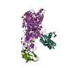
| ||||||||||||
|---|---|---|---|---|---|---|---|---|---|---|---|---|---|
| 1 | 
| ||||||||||||
| Unit cell |
| ||||||||||||
| Components on special symmetry positions |
|
-Components
-Urease subunit ... , 3 types, 3 molecules AAABBBCCC
| #1: Protein | Mass: 11134.895 Da / Num. of mol.: 1 / Source method: isolated from a natural source / Source: (natural) Sporosarcina pasteurii (bacteria) / References: UniProt: P41022, urease |
|---|---|
| #2: Protein | Mass: 13529.061 Da / Num. of mol.: 1 / Source method: isolated from a natural source / Source: (natural) Sporosarcina pasteurii (bacteria) / References: UniProt: P41021, urease |
| #3: Protein | Mass: 61575.648 Da / Num. of mol.: 1 / Source method: isolated from a natural source / Source: (natural) Sporosarcina pasteurii (bacteria) / References: UniProt: P41020, urease |
-Non-polymers , 6 types, 501 molecules 


| #4: Chemical | ChemComp-EDO /  Mass: 62.068 Da / Num. of mol.: 13 / Source method: obtained synthetically / Formula: C2H6O2 Mass: 62.068 Da / Num. of mol.: 13 / Source method: obtained synthetically / Formula: C2H6O2#5: Chemical | ChemComp-SO4 /  Mass: 96.063 Da / Num. of mol.: 10 / Source method: obtained synthetically / Formula: SO4 Mass: 96.063 Da / Num. of mol.: 10 / Source method: obtained synthetically / Formula: SO4#6: Chemical |  Mass: 58.693 Da / Num. of mol.: 2 / Source method: obtained synthetically / Formula: Ni Mass: 58.693 Da / Num. of mol.: 2 / Source method: obtained synthetically / Formula: Ni#7: Chemical | ChemComp-O / |  Mass: 15.999 Da / Num. of mol.: 1 / Source method: obtained synthetically / Formula: O Mass: 15.999 Da / Num. of mol.: 1 / Source method: obtained synthetically / Formula: O#8: Chemical |  Mass: 107.868 Da / Num. of mol.: 2 / Source method: obtained synthetically / Formula: Ag / Feature type: SUBJECT OF INVESTIGATION Mass: 107.868 Da / Num. of mol.: 2 / Source method: obtained synthetically / Formula: Ag / Feature type: SUBJECT OF INVESTIGATION#9: Water | ChemComp-HOH / | Mass: 18.015 Da / Num. of mol.: 473 / Source method: isolated from a natural source / Formula: H2O |
|---|
-Details
| Has ligand of interest | Y |
|---|
-Experimental details
-Experiment
| Experiment | Method: X-RAY DIFFRACTION / Number of used crystals: 1 |
|---|
- Sample preparation
| Crystal | Density Matthews: 2.74 Å3/Da / Density % sol: 55.14 % / Description: Rice-shaped |
|---|---|
| Crystal grow | Temperature: 293 K / Method: vapor diffusion, hanging drop / pH: 6.5 Details: THE PROTEIN-LIGAND (300 microM) COMPLEX IN 50 mM HEPES BUFFER, PH 7.50 (ALSO CONTAINING 1% (V/V) DMSO), DILUTED 1:1 WITH A SOLUTION OF 1.4 M AMMONIUM SULFATE ALSO CONTAINING THE SAME ...Details: THE PROTEIN-LIGAND (300 microM) COMPLEX IN 50 mM HEPES BUFFER, PH 7.50 (ALSO CONTAINING 1% (V/V) DMSO), DILUTED 1:1 WITH A SOLUTION OF 1.4 M AMMONIUM SULFATE ALSO CONTAINING THE SAME CONCENTRATION OF LIGAND AND DMSO. PH range: 6.3-6.8 |
-Data collection
| Diffraction | Mean temperature: 100 K / Serial crystal experiment: N |
|---|---|
| Diffraction source | Source: SYNCHROTRON / Site: PETRA III, EMBL c/o DESY  / Beamline: P13 (MX1) / Wavelength: 0.9762 Å / Beamline: P13 (MX1) / Wavelength: 0.9762 Å |
| Detector | Type: DECTRIS PILATUS 6M / Detector: PIXEL / Date: Nov 30, 2019 |
| Radiation | Monochromator: SI (111) / Protocol: SINGLE WAVELENGTH / Monochromatic (M) / Laue (L): M / Scattering type: x-ray |
| Radiation wavelength | Wavelength: 0.9762 Å / Relative weight: 1 |
| Reflection | Resolution: 1.97→113.87 Å / Num. obs: 68762 / % possible obs: 100 % / Redundancy: 13.2 % / Biso Wilson estimate: 29.2 Å2 / CC1/2: 0.998 / Rmerge(I) obs: 0.2 / Rpim(I) all: 0.064 / Rrim(I) all: 0.234 / Net I/σ(I): 12.2 |
| Reflection shell | Resolution: 1.97→2.02 Å / Redundancy: 12.4 % / Rmerge(I) obs: 3.191 / Mean I/σ(I) obs: 1 / Num. unique obs: 4556 / CC1/2: 0.593 / Rpim(I) all: 0.982 / Rrim(I) all: 3.473 / % possible all: 99.9 |
- Processing
| Software |
| |||||||||||||||||||||||||||||||||||||||||||||||||||||||||||||||||||||||||||||||||||||||||||||||||||||||||||||||||||||||||||||||||||||||||||||||||||||||||||
|---|---|---|---|---|---|---|---|---|---|---|---|---|---|---|---|---|---|---|---|---|---|---|---|---|---|---|---|---|---|---|---|---|---|---|---|---|---|---|---|---|---|---|---|---|---|---|---|---|---|---|---|---|---|---|---|---|---|---|---|---|---|---|---|---|---|---|---|---|---|---|---|---|---|---|---|---|---|---|---|---|---|---|---|---|---|---|---|---|---|---|---|---|---|---|---|---|---|---|---|---|---|---|---|---|---|---|---|---|---|---|---|---|---|---|---|---|---|---|---|---|---|---|---|---|---|---|---|---|---|---|---|---|---|---|---|---|---|---|---|---|---|---|---|---|---|---|---|---|---|---|---|---|---|---|---|---|
| Refinement | Method to determine structure: MOLECULAR REPLACEMENT Starting model: 6G48 Resolution: 1.97→97.798 Å / Cor.coef. Fo:Fc: 0.975 / Cor.coef. Fo:Fc free: 0.958 / SU B: 5.99 / SU ML: 0.143 / Cross valid method: FREE R-VALUE / ESU R: 0.141 / ESU R Free: 0.135 Details: Hydrogens have been added in their riding positions
| |||||||||||||||||||||||||||||||||||||||||||||||||||||||||||||||||||||||||||||||||||||||||||||||||||||||||||||||||||||||||||||||||||||||||||||||||||||||||||
| Solvent computation | Ion probe radii: 0.8 Å / Shrinkage radii: 0.8 Å / VDW probe radii: 1.2 Å / Solvent model: MASK BULK SOLVENT | |||||||||||||||||||||||||||||||||||||||||||||||||||||||||||||||||||||||||||||||||||||||||||||||||||||||||||||||||||||||||||||||||||||||||||||||||||||||||||
| Displacement parameters | Biso mean: 37.581 Å2
| |||||||||||||||||||||||||||||||||||||||||||||||||||||||||||||||||||||||||||||||||||||||||||||||||||||||||||||||||||||||||||||||||||||||||||||||||||||||||||
| Refinement step | Cycle: LAST / Resolution: 1.97→97.798 Å
| |||||||||||||||||||||||||||||||||||||||||||||||||||||||||||||||||||||||||||||||||||||||||||||||||||||||||||||||||||||||||||||||||||||||||||||||||||||||||||
| Refine LS restraints |
| |||||||||||||||||||||||||||||||||||||||||||||||||||||||||||||||||||||||||||||||||||||||||||||||||||||||||||||||||||||||||||||||||||||||||||||||||||||||||||
| LS refinement shell |
|
