 Movie
Movie Controller


+ Open data

- Basic information
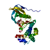
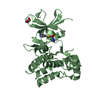
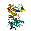
| Entry | Database: PDB / ID: 7b3a | ||||||
|---|---|---|---|---|---|---|---|
| Title | Crystal structure of PamZ | ||||||
 Components Components | Putative Acyl-transferase domain protein | ||||||
 Keywords Keywords | TRANSFERASE / GCN5-RELATED N-ACETYLTRANSFERASE-LIKE PROTEIN / paenilamicin / paenilamicin-gene cluster | ||||||
| Function / homology | GNAT acetyltransferase YdfB-like / GNAT acetyltransferase / N-acetyltransferase activity / Gcn5-related N-acetyltransferase (GNAT) domain profile. / GNAT domain / Acyl-CoA N-acyltransferase / ACETYL COENZYME *A / ACETATE ION / Putative Acyl-transferase domain protein Function and homology information Function and homology information | ||||||
| Biological species |  Paenibacillus larvae subsp. larvae (bacteria) Paenibacillus larvae subsp. larvae (bacteria) | ||||||
| Method |  X-RAY DIFFRACTION / SYNCHROTRON / MOLECULAR REPLACEMENT / Resolution: 1.34 Å X-RAY DIFFRACTION / SYNCHROTRON / MOLECULAR REPLACEMENT / Resolution: 1.34 Å | ||||||
 Authors Authors | Loll, B. / Dang, T. / Mainz, A. / Suessmuth, R. / Wahl, M.C. | ||||||
 Citation Citation | Journal: Nat Commun / Year: 2022 Title: Molecular basis of antibiotic self-resistance in a bee larvae pathogen. Authors: Dang, T. / Loll, B. / Muller, S. / Skobalj, R. / Ebeling, J. / Bulatov, T. / Gensel, S. / Gobel, J. / Wahl, M.C. / Genersch, E. / Mainz, A. / Sussmuth, R.D. | ||||||
| History |
|
- Structure visualization
| Structure viewer | Molecule: MolmilJmol/JSmol |
|---|
- Downloads & links
-Download
| PDBx/mmCIF format | 7b3a.cif.gz | 238.8 KB | Display | PDBx/mmCIF format |
|---|---|---|---|---|
| PDB format | pdb7b3a.ent.gz | 161.9 KB | Display | PDB format |
| PDBx/mmJSON format | 7b3a.json.gz | Tree view | PDBx/mmJSON format | |
| Others |  Other downloads Other downloads |
-Validation report
| Summary document | 7b3a_validation.pdf.gz | 439 KB | Display | wwPDB validaton report |
|---|---|---|---|---|
| Full document | 7b3a_full_validation.pdf.gz | 440 KB | Display | |
| Data in XML | 7b3a_validation.xml.gz | 14.9 KB | Display | |
| Data in CIF | 7b3a_validation.cif.gz | 23 KB | Display | |
| Arichive directory | https://data.pdbj.org/pub/pdb/validation_reports/b3/7b3aftp://data.pdbj.org/pub/pdb/validation_reports/b3/7b3a | HTTPS FTP |
-Related structure data
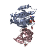
| Related structure data | 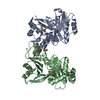 3g3sS S: Starting model for refinement |
|---|---|
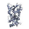
| Similar structure data |


-Links
 PDBj
PDBj

- Assembly
| Deposited unit | 
| ||||||||||||
|---|---|---|---|---|---|---|---|---|---|---|---|---|---|
| 1 |
| ||||||||||||
| Unit cell |
|
-Components
| #1: Protein | Mass: 32271.830 Da / Num. of mol.: 1 Source method: isolated from a genetically manipulated source Source: (gene. exp.) Paenibacillus larvae subsp. larvae (bacteria)Gene: ERICIII_02339, ERICV_02787 / Plasmid: pet28 / Production host: |
|---|---|
| #2: Chemical | ChemComp-ACO /   Mass: 809.571 Da / Num. of mol.: 1 / Source method: obtained synthetically / Formula: C23H38N7O17P3S Mass: 809.571 Da / Num. of mol.: 1 / Source method: obtained synthetically / Formula: C23H38N7O17P3S |
| #3: Chemical | ChemComp-CL /   Mass: 35.453 Da / Num. of mol.: 1 / Source method: obtained synthetically / Formula: Cl Mass: 35.453 Da / Num. of mol.: 1 / Source method: obtained synthetically / Formula: Cl |
| #4: Chemical | ChemComp-ACT /   Mass: 59.044 Da / Num. of mol.: 1 / Source method: obtained synthetically / Formula: C2H3O2 Mass: 59.044 Da / Num. of mol.: 1 / Source method: obtained synthetically / Formula: C2H3O2 |
| #5: Water | ChemComp-HOH /  Mass: 18.015 Da / Num. of mol.: 270 / Source method: isolated from a natural source / Formula: H2O Mass: 18.015 Da / Num. of mol.: 270 / Source method: isolated from a natural source / Formula: H2O |
| Has ligand of interest | N |
-Experimental details
-Experiment
| Experiment | Method: X-RAY DIFFRACTION / Number of used crystals: 1 |
|---|
- Sample preparation
| Crystal | Density Matthews: 2.1 Å3/Da / Density % sol: 41.3 % |
|---|---|
| Crystal grow | Temperature: 293 K / Method: vapor diffusion, sitting drop / pH: 4.6 Details: 40% (w/v) PEG 3350, 50 mM ammonium sulfate, 100 mM sodium acetate at pH 4.6. |
-Data collection
| Diffraction | Mean temperature: 100 K / Serial crystal experiment: N |
|---|---|
| Diffraction source | Source: SYNCHROTRON / Site: BESSY  / Beamline: 14.1 / Wavelength: 0.941841 Å / Beamline: 14.1 / Wavelength: 0.941841 Å |
| Detector | Type: DECTRIS PILATUS 6M / Detector: PIXEL / Date: Dec 13, 2018 |
| Radiation | Protocol: SINGLE WAVELENGTH / Monochromatic (M) / Laue (L): M / Scattering type: x-ray |
| Radiation wavelength | Wavelength: 0.941841 Å / Relative weight: 1 |
| Reflection | Resolution: 1.34→50 Å / Num. obs: 58898 / % possible obs: 98.2 % / Redundancy: 3.8 % / Biso Wilson estimate: 16.48 Å2 / CC1/2: 0.999 / Rrim(I) all: 0.068 / Net I/σ(I): 11.2 |
| Reflection shell | Resolution: 1.34→1.42 Å / Mean I/σ(I) obs: 1 / Num. unique obs: 9129 / CC1/2: 0.379 / Rrim(I) all: 129.2 |
- Processing
| Software |
| ||||||||||||||||||||||||||||||||||||||||||||||||||||||||||||||||||||||||||||||||||||||||||||||||||||||||||||||||
|---|---|---|---|---|---|---|---|---|---|---|---|---|---|---|---|---|---|---|---|---|---|---|---|---|---|---|---|---|---|---|---|---|---|---|---|---|---|---|---|---|---|---|---|---|---|---|---|---|---|---|---|---|---|---|---|---|---|---|---|---|---|---|---|---|---|---|---|---|---|---|---|---|---|---|---|---|---|---|---|---|---|---|---|---|---|---|---|---|---|---|---|---|---|---|---|---|---|---|---|---|---|---|---|---|---|---|---|---|---|---|---|---|---|
| Refinement | Method to determine structure: MOLECULAR REPLACEMENT Starting model: 3G3S Resolution: 1.34→20.2 Å / SU ML: 0.166 / Cross valid method: FREE R-VALUE / σ(F): 1.36 / Phase error: 20.038 Stereochemistry target values: GeoStd + Monomer Library + CDL v1.2
| ||||||||||||||||||||||||||||||||||||||||||||||||||||||||||||||||||||||||||||||||||||||||||||||||||||||||||||||||
| Solvent computation | Shrinkage radii: 0.9 Å / VDW probe radii: 1.11 Å / Solvent model: FLAT BULK SOLVENT MODEL | ||||||||||||||||||||||||||||||||||||||||||||||||||||||||||||||||||||||||||||||||||||||||||||||||||||||||||||||||
| Displacement parameters | Biso mean: 24.5 Å2 | ||||||||||||||||||||||||||||||||||||||||||||||||||||||||||||||||||||||||||||||||||||||||||||||||||||||||||||||||
| Refinement step | Cycle: LAST / Resolution: 1.34→20.2 Å
| ||||||||||||||||||||||||||||||||||||||||||||||||||||||||||||||||||||||||||||||||||||||||||||||||||||||||||||||||
| Refine LS restraints |
| ||||||||||||||||||||||||||||||||||||||||||||||||||||||||||||||||||||||||||||||||||||||||||||||||||||||||||||||||
| LS refinement shell |
|
