 Movie
Movie Controller

[English] 日本語
 Yorodumi
- PDB-7b0p: In meso structure of the membrane integral lipoprotein intramolec... -
+ Open data

- Basic information
| Entry | Database: PDB / ID: 7b0p | ||||||
|---|---|---|---|---|---|---|---|
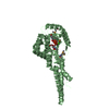
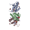
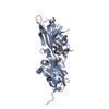
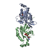
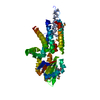
| Title | In meso structure of the membrane integral lipoprotein intramolecular transacylase Lit from Bacillus cereus in space group P21212 | ||||||
 Components Components | Hypothetical Membrane Spanning Protein | ||||||
 Keywords Keywords | MEMBRANE PROTEIN / lipoprotein / lipid cubic phase / Lit / transacylase | ||||||
| Function / homology | Integral membrane protein 1906 / Lipoprotein intramolecular transacylase Lit / (2R)-2,3-dihydroxypropyl (9Z)-octadec-9-enoate / 1-METHOXY-2-[2-(2-METHOXY-ETHOXY]-ETHANE / Hypothetical Membrane Spanning Protein Function and homology information Function and homology information | ||||||
| Biological species |  | ||||||
| Method |  X-RAY DIFFRACTION / SYNCHROTRON / MOLECULAR REPLACEMENT / Resolution: 1.935 Å X-RAY DIFFRACTION / SYNCHROTRON / MOLECULAR REPLACEMENT / Resolution: 1.935 Å | ||||||
 Authors Authors | Huang, C.-Y. / Olatunji, S. / Olieric, V. / Caffrey, M. | ||||||
| Funding support |  Ireland, 1items Ireland, 1items
| ||||||
 Citation Citation | Journal: Nat Commun / Year: 2021 Title: Structural basis of the membrane intramolecular transacylase reaction responsible for lyso-form lipoprotein synthesis. Authors: Olatunji, S. / Bowen, K. / Huang, C.Y. / Weichert, D. / Singh, W. / Tikhonova, I.G. / Scanlan, E.M. / Olieric, V. / Caffrey, M. | ||||||
| History |
|
- Structure visualization
| Structure viewer | Molecule: MolmilJmol/JSmol |
|---|
- Downloads & links
-Download
| PDBx/mmCIF format | 7b0p.cif.gz | 215.9 KB | Display | PDBx/mmCIF format |
|---|---|---|---|---|
| PDB format | pdb7b0p.ent.gz | 174.5 KB | Display | PDB format |
| PDBx/mmJSON format | 7b0p.json.gz | Tree view | PDBx/mmJSON format | |
| Others |  Other downloads Other downloads |
-Validation report
| Arichive directory | https://data.pdbj.org/pub/pdb/validation_reports/b0/7b0pftp://data.pdbj.org/pub/pdb/validation_reports/b0/7b0p | HTTPS FTP |
|---|
-Related structure data
| Related structure data | 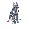 7b0oSC 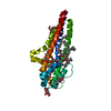 7b0qC 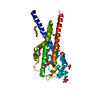 7b0rC S: Starting model for refinement C: citing same article ( |
|---|---|
| Similar structure data |

-Links
 PDBj
PDBj


- Assembly
| Deposited unit | 
| |||||||||||||||||||||||||||||||||||||||||||||||||||
|---|---|---|---|---|---|---|---|---|---|---|---|---|---|---|---|---|---|---|---|---|---|---|---|---|---|---|---|---|---|---|---|---|---|---|---|---|---|---|---|---|---|---|---|---|---|---|---|---|---|---|---|---|
| 1 | 
| |||||||||||||||||||||||||||||||||||||||||||||||||||
| 2 | 
| |||||||||||||||||||||||||||||||||||||||||||||||||||
| Unit cell |
| |||||||||||||||||||||||||||||||||||||||||||||||||||
| Noncrystallographic symmetry (NCS) | NCS domain:
NCS domain segments:
|
-Components
| #1: Protein | Mass: 27640.795 Da / Num. of mol.: 2 Source method: isolated from a genetically manipulated source Source: (gene. exp.) Strain: ATCC 14579 / DSM 31 / JCM 2152 / NBRC 15305 / NCIMB 9373 / NRRL B-3711 Gene: BC_1526 / Production host: #2: Chemical | ChemComp-OLC / (   Mass: 356.540 Da / Num. of mol.: 12 / Source method: obtained synthetically / Formula: C21H40O4 / Feature type: SUBJECT OF INVESTIGATION Mass: 356.540 Da / Num. of mol.: 12 / Source method: obtained synthetically / Formula: C21H40O4 / Feature type: SUBJECT OF INVESTIGATION#3: Chemical | ChemComp-GOL /   Mass: 92.094 Da / Num. of mol.: 15 / Source method: obtained synthetically / Formula: C3H8O3 Mass: 92.094 Da / Num. of mol.: 15 / Source method: obtained synthetically / Formula: C3H8O3#4: Chemical | ChemComp-PG5 / |   Mass: 178.226 Da / Num. of mol.: 1 / Source method: obtained synthetically / Formula: C8H18O4 Mass: 178.226 Da / Num. of mol.: 1 / Source method: obtained synthetically / Formula: C8H18O4#5: Water | ChemComp-HOH / |  Mass: 18.015 Da / Num. of mol.: 168 / Source method: isolated from a natural source / Formula: H2O Mass: 18.015 Da / Num. of mol.: 168 / Source method: isolated from a natural source / Formula: H2OHas ligand of interest | Y | |
|---|
-Experimental details
-Experiment
| Experiment | Method: X-RAY DIFFRACTION / Number of used crystals: 1 |
|---|
- Sample preparation
| Crystal | Density Matthews: 3.42 Å3/Da / Density % sol: 63.99 % |
|---|---|
| Crystal grow | Temperature: 293 K / Method: lipidic cubic phase / pH: 5.5 Details: 100 mM sodium citrate/HCl, pH 5.5, 75-150 mM NaCl, and 36-44 %(v/v) PEG 200 |
-Data collection
| Diffraction | Mean temperature: 100 K / Serial crystal experiment: N |
|---|---|
| Diffraction source | Source: SYNCHROTRON / Site: Diamond  / Beamline: I24 / Wavelength: 0.9686 Å / Beamline: I24 / Wavelength: 0.9686 Å |
| Detector | Type: DECTRIS PILATUS3 6M / Detector: PIXEL / Date: Mar 8, 2019 |
| Radiation | Protocol: SINGLE WAVELENGTH / Monochromatic (M) / Laue (L): M / Scattering type: x-ray |
| Radiation wavelength | Wavelength: 0.9686 Å / Relative weight: 1 |
| Reflection | Resolution: 1.935→60 Å / Num. obs: 29001 / % possible obs: 89.5 % / Redundancy: 5.4 % / CC1/2: 0.73 / Rrim(I) all: 0.66 / Net I/σ(I): 4.4 |
| Reflection shell | Resolution: 1.94→2.28 Å / Mean I/σ(I) obs: 1.7 / Num. unique obs: 1451 / CC1/2: 0.162 / Rrim(I) all: 6.07 / % possible all: 47.4 |
- Processing
| Software |
| ||||||||||||||||||||||||||||||||||||||||||||||||||||||||||||||||||
|---|---|---|---|---|---|---|---|---|---|---|---|---|---|---|---|---|---|---|---|---|---|---|---|---|---|---|---|---|---|---|---|---|---|---|---|---|---|---|---|---|---|---|---|---|---|---|---|---|---|---|---|---|---|---|---|---|---|---|---|---|---|---|---|---|---|---|---|
| Refinement | Method to determine structure: MOLECULAR REPLACEMENT Starting model: 7B0O Resolution: 1.935→60 Å / SU ML: 0.21 / Cross valid method: THROUGHOUT / σ(F): 1.33 / Phase error: 29.01 / Stereochemistry target values: ML
| ||||||||||||||||||||||||||||||||||||||||||||||||||||||||||||||||||
| Solvent computation | Shrinkage radii: 0.9 Å / VDW probe radii: 1.11 Å / Solvent model: FLAT BULK SOLVENT MODEL | ||||||||||||||||||||||||||||||||||||||||||||||||||||||||||||||||||
| Displacement parameters | Biso max: 149.08 Å2 / Biso mean: 34.3226 Å2 / Biso min: 3.84 Å2 | ||||||||||||||||||||||||||||||||||||||||||||||||||||||||||||||||||
| Refinement step | Cycle: final / Resolution: 1.935→60 Å
| ||||||||||||||||||||||||||||||||||||||||||||||||||||||||||||||||||
| Refine LS restraints NCS |
| ||||||||||||||||||||||||||||||||||||||||||||||||||||||||||||||||||
| LS refinement shell | Refine-ID: X-RAY DIFFRACTION / Rfactor Rfree error: 0
| ||||||||||||||||||||||||||||||||||||||||||||||||||||||||||||||||||
| Refinement TLS params. | Method: refined / Origin x: -18.3228 Å / Origin y: 28.5323 Å / Origin z: -28.3655 Å
| ||||||||||||||||||||||||||||||||||||||||||||||||||||||||||||||||||
| Refinement TLS group |
|
