 Movie
Movie Controller


+ Open data

- Basic information
| Entry | Database: PDB / ID: 7axu | ||||||
|---|---|---|---|---|---|---|---|
| Title | Structure of WDR5:CS-VIP8 cocrystal after illumination in situ | ||||||
 Components Components | WD repeat-containing protein 5 | ||||||
 Keywords Keywords | TRANSFERASE / WDR5 / cyclic strained visible-light photoswitches / MLL1 complex disruption / inhibition of hematopoiesis | ||||||
| Function / homology |  Function and homology information Function and homology informationhistone H3Q5ser reader activity / histone H3K4me1 reader activity / Loss of Function of KMT2D in MLL4 Complex Formation in Kabuki Syndrome / Epigenetic regulation of gene expression by MLL3 and MLL4 complexes / MLL3/4 complex / Set1C/COMPASS complex / ATAC complex / MLL1/2 complex / NSL complex / histone H3K4 methyltransferase activity ...histone H3Q5ser reader activity / histone H3K4me1 reader activity / Loss of Function of KMT2D in MLL4 Complex Formation in Kabuki Syndrome / Epigenetic regulation of gene expression by MLL3 and MLL4 complexes / MLL3/4 complex / Set1C/COMPASS complex / ATAC complex / MLL1/2 complex / NSL complex / histone H3K4 methyltransferase activity / Cardiogenesis / Formation of WDR5-containing histone-modifying complexes / histone methyltransferase complex / MLL1 complex / regulation of embryonic development / histone acetyltransferase complex / regulation of cell division / positive regulation of gluconeogenesis / transcription initiation-coupled chromatin remodeling / gluconeogenesis / skeletal system development / RUNX1 regulates genes involved in megakaryocyte differentiation and platelet function / PKMTs methylate histone lysines / Activation of anterior HOX genes in hindbrain development during early embryogenesis / RMTs methylate histone arginines / mitotic spindle / HATs acetylate histones / MLL4 and MLL3 complexes regulate expression of PPARG target genes in adipogenesis and hepatic steatosis / sperm principal piece / Neddylation / histone binding / regulation of cell cycle / regulation of transcription by RNA polymerase II / regulation of DNA-templated transcription / positive regulation of DNA-templated transcription / negative regulation of transcription by RNA polymerase II / nucleoplasm / nucleus Similarity search - Function | ||||||
| Biological species |  Homo sapiens (human) Homo sapiens (human) | ||||||
| Method |  X-RAY DIFFRACTION / SYNCHROTRON / MOLECULAR REPLACEMENT / Resolution: 1.68 Å X-RAY DIFFRACTION / SYNCHROTRON / MOLECULAR REPLACEMENT / Resolution: 1.68 Å | ||||||
 Authors Authors | Werel, L. / Essen, L.-O. | ||||||
 Citation Citation | Journal: Acs Cent.Sci. / Year: 2022 Title: Bistable Photoswitch Allows in Vivo Control of Hematopoiesis. Authors: Albert, L. / Nagpal, J. / Steinchen, W. / Zhang, L. / Werel, L. / Djokovic, N. / Ruzic, D. / Hoffarth, M. / Xu, J. / Kaspareit, J. / Abendroth, F. / Royant, A. / Bange, G. / Nikolic, K. / ...Authors: Albert, L. / Nagpal, J. / Steinchen, W. / Zhang, L. / Werel, L. / Djokovic, N. / Ruzic, D. / Hoffarth, M. / Xu, J. / Kaspareit, J. / Abendroth, F. / Royant, A. / Bange, G. / Nikolic, K. / Ryu, S. / Dou, Y. / Essen, L.O. / Vazquez, O. | ||||||
| History |
|
- Structure visualization
| Structure viewer | Molecule: MolmilJmol/JSmol |
|---|
- Downloads & links
-Download
| PDBx/mmCIF format | 7axu.cif.gz | 84.3 KB | Display | PDBx/mmCIF format |
|---|---|---|---|---|
| PDB format | pdb7axu.ent.gz | 61.2 KB | Display | PDB format |
| PDBx/mmJSON format | 7axu.json.gz | Tree view | PDBx/mmJSON format | |
| Others |  Other downloads Other downloads |
-Validation report
| Arichive directory | https://data.pdbj.org/pub/pdb/validation_reports/ax/7axuftp://data.pdbj.org/pub/pdb/validation_reports/ax/7axu | HTTPS FTP |
|---|
-Related structure data
| Related structure data |  7axpC 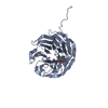 7axqC 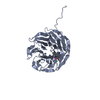 7axsC  7axxC 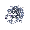 6iamS C: citing same article ( S: Starting model for refinement |
|---|---|
| Similar structure data |


-Links
 PDBj
PDBj- Assembly
| Deposited unit | 
| ||||||||
|---|---|---|---|---|---|---|---|---|---|
| 1 |
| ||||||||
| Unit cell |
|
-Components
| #1: Protein | Mass: 36635.438 Da / Num. of mol.: 1 Source method: isolated from a genetically manipulated source Details: WDR5, cyclic strained visible-light photoswitches, MLL1 complex disruption, inhibition of hematopoiesis Source: (gene. exp.) Homo sapiens (human) / Gene: WDR5, BIG3 / Production host:  |
|---|---|
| #2: Water | ChemComp-HOH /  Mass: 18.015 Da / Num. of mol.: 392 / Source method: isolated from a natural source / Formula: H2O Mass: 18.015 Da / Num. of mol.: 392 / Source method: isolated from a natural source / Formula: H2O |
-Experimental details
-Experiment
| Experiment | Method: X-RAY DIFFRACTION / Number of used crystals: 1 |
|---|
- Sample preparation
| Crystal | Density Matthews: 2.04 Å3/Da / Density % sol: 39.6 % |
|---|---|
| Crystal grow | Temperature: 277.15 K / Method: vapor diffusion, sitting drop / pH: 8.5 Details: 10% (w/v) PEG20000, 20% (v/v) PEG550 MME, 0.02 M sodium formate, 0.02 M ammonium acetate, 0.02 M trisodium citrate, 0.02 M sodium potassium L-tartrate, 0.02 M sodium oxamate |
-Data collection
| Diffraction | Mean temperature: 100 K / Serial crystal experiment: N |
|---|---|
| Diffraction source | Source: SYNCHROTRON / Site: SLS  / Beamline: X06SA / Wavelength: 1 Å / Beamline: X06SA / Wavelength: 1 Å |
| Detector | Type: DECTRIS EIGER X 16M / Detector: PIXEL / Date: Aug 19, 2019 |
| Radiation | Protocol: SINGLE WAVELENGTH / Monochromatic (M) / Laue (L): M / Scattering type: x-ray |
| Radiation wavelength | Wavelength: 1 Å / Relative weight: 1 |
| Reflection | Resolution: 1.68→44.7 Å / Num. obs: 34930 / % possible obs: 99.84 % / Redundancy: 2 % / Biso Wilson estimate: 16.1 Å2 / CC1/2: 0.998 / Rmerge(I) obs: 0.02835 / Rpim(I) all: 0.02835 / Rrim(I) all: 0.04009 / Net I/σ(I): 15.39 |
| Reflection shell | Resolution: 1.68→1.74 Å / Redundancy: 2 % / Rmerge(I) obs: 0.1981 / Mean I/σ(I) obs: 3.5 / Num. unique obs: 6864 / CC1/2: 0.922 / Rpim(I) all: 0.1981 / Rrim(I) all: 0.2802 / % possible all: 99.8 |
- Processing
| Software |
| |||||||||||||||||||||||||||||||||||||||||||||||||||||||||||||||||||||||||||||||||||||||||||||||||||||||||
|---|---|---|---|---|---|---|---|---|---|---|---|---|---|---|---|---|---|---|---|---|---|---|---|---|---|---|---|---|---|---|---|---|---|---|---|---|---|---|---|---|---|---|---|---|---|---|---|---|---|---|---|---|---|---|---|---|---|---|---|---|---|---|---|---|---|---|---|---|---|---|---|---|---|---|---|---|---|---|---|---|---|---|---|---|---|---|---|---|---|---|---|---|---|---|---|---|---|---|---|---|---|---|---|---|---|---|
| Refinement | Method to determine structure: MOLECULAR REPLACEMENT Starting model: 6IAM Resolution: 1.68→44.7 Å / Cor.coef. Fo:Fc: 0.964 / Cor.coef. Fo:Fc free: 0.952 / SU B: 1.81 / SU ML: 0.06 / Cross valid method: THROUGHOUT / ESU R: 0.098 / ESU R Free: 0.097 / Stereochemistry target values: MAXIMUM LIKELIHOOD
| |||||||||||||||||||||||||||||||||||||||||||||||||||||||||||||||||||||||||||||||||||||||||||||||||||||||||
| Solvent computation | Ion probe radii: 0.7 Å / Shrinkage radii: 0.7 Å / VDW probe radii: 1 Å / Solvent model: MASK | |||||||||||||||||||||||||||||||||||||||||||||||||||||||||||||||||||||||||||||||||||||||||||||||||||||||||
| Displacement parameters | Biso mean: 17.39 Å2
| |||||||||||||||||||||||||||||||||||||||||||||||||||||||||||||||||||||||||||||||||||||||||||||||||||||||||
| Refinement step | Cycle: LAST / Resolution: 1.68→44.7 Å
| |||||||||||||||||||||||||||||||||||||||||||||||||||||||||||||||||||||||||||||||||||||||||||||||||||||||||
| Refine LS restraints |
| |||||||||||||||||||||||||||||||||||||||||||||||||||||||||||||||||||||||||||||||||||||||||||||||||||||||||
| LS refinement shell | Resolution: 1.68→1.723 Å
|
