 Movie
Movie Controller

[English] 日本語
 Yorodumi
- PDB-7ax2: Crystal structure of the computationally designed Scone-E protein... -
+ Open data

- Basic information
| Entry | Database: PDB / ID: 7ax2 | |||||||||||||||
|---|---|---|---|---|---|---|---|---|---|---|---|---|---|---|---|---|
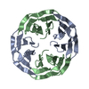
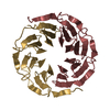
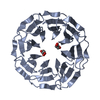
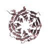
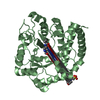
| Title | Crystal structure of the computationally designed Scone-E protein co-crystallized with STA, form b | |||||||||||||||
 Components Components | Scone-E | |||||||||||||||
 Keywords Keywords | DE NOVO PROTEIN / Beta-propeller / Computational design | |||||||||||||||
| Function / homology | Monolacunary Keggin (STA) / Keggin (STA) Function and homology information Function and homology information | |||||||||||||||
| Biological species | synthetic construct (others) | |||||||||||||||
| Method |  X-RAY DIFFRACTION / SYNCHROTRON / MOLECULAR REPLACEMENT / Resolution: 2.1 Å X-RAY DIFFRACTION / SYNCHROTRON / MOLECULAR REPLACEMENT / Resolution: 2.1 Å | |||||||||||||||
 Authors Authors | Mylemans, B. / Vandebroek, L. / Parac-Vogt, T.N. / Voet, A.R.D. | |||||||||||||||
| Funding support |  Belgium, 4items Belgium, 4items
| |||||||||||||||
 Citation Citation | Journal: Acta Crystallogr D Struct Biol / Year: 2021 Title: Crystal structures of Scone: pseudosymmetric folding of a symmetric designer protein. Authors: Mylemans, B. / Killian, T. / Vandebroek, L. / Van Meervelt, L. / Tame, J.R.H. / Parac-Vogt, T.N. / Voet, A.R.D. | |||||||||||||||
| History |
|
- Structure visualization
| Structure viewer | Molecule: MolmilJmol/JSmol |
|---|
- Downloads & links
-Download
| PDBx/mmCIF format | 7ax2.cif.gz | 79.6 KB | Display | PDBx/mmCIF format |
|---|---|---|---|---|
| PDB format | pdb7ax2.ent.gz | 55.8 KB | Display | PDB format |
| PDBx/mmJSON format | 7ax2.json.gz | Tree view | PDBx/mmJSON format | |
| Others |  Other downloads Other downloads |
-Validation report
| Summary document | 7ax2_validation.pdf.gz | 1.8 MB | Display | wwPDB validaton report |
|---|---|---|---|---|
| Full document | 7ax2_full_validation.pdf.gz | 1.8 MB | Display | |
| Data in XML | 7ax2_validation.xml.gz | 13.9 KB | Display | |
| Data in CIF | 7ax2_validation.cif.gz | 19.1 KB | Display | |
| Arichive directory | https://data.pdbj.org/pub/pdb/validation_reports/ax/7ax2ftp://data.pdbj.org/pub/pdb/validation_reports/ax/7ax2 | HTTPS FTP |
-Related structure data
| Related structure data |  7awyC  7awzC 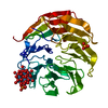 7ax0C  6tjgS S: Starting model for refinement C: citing same article ( |
|---|---|
| Similar structure data |


-Links
 PDBj
PDBj
- Assembly
| Deposited unit | 
| ||||||||||||
|---|---|---|---|---|---|---|---|---|---|---|---|---|---|
| 1 |
| ||||||||||||
| Unit cell |
|
-Components
| #1: Protein | Mass: 40116.891 Da / Num. of mol.: 1 Source method: isolated from a genetically manipulated source Source: (gene. exp.) synthetic construct (others) / Production host:  |
|---|---|
| #2: Chemical | ChemComp-SIW /   Mass: 2874.142 Da / Num. of mol.: 1 / Source method: obtained synthetically / Formula: O40SiW12 / Feature type: SUBJECT OF INVESTIGATION Mass: 2874.142 Da / Num. of mol.: 1 / Source method: obtained synthetically / Formula: O40SiW12 / Feature type: SUBJECT OF INVESTIGATION |
| #3: Chemical | ChemComp-S5T /   Mass: 2674.302 Da / Num. of mol.: 1 / Source method: obtained synthetically / Formula: O39SiW11 / Feature type: SUBJECT OF INVESTIGATION Mass: 2674.302 Da / Num. of mol.: 1 / Source method: obtained synthetically / Formula: O39SiW11 / Feature type: SUBJECT OF INVESTIGATION |
| #4: Chemical | ChemComp-NA /   Mass: 22.990 Da / Num. of mol.: 1 / Source method: obtained synthetically / Formula: Na Mass: 22.990 Da / Num. of mol.: 1 / Source method: obtained synthetically / Formula: Na |
| #5: Water | ChemComp-HOH /  Mass: 18.015 Da / Num. of mol.: 79 / Source method: isolated from a natural source / Formula: H2O Mass: 18.015 Da / Num. of mol.: 79 / Source method: isolated from a natural source / Formula: H2O |
| Has ligand of interest | Y |
-Experimental details
-Experiment
| Experiment | Method: X-RAY DIFFRACTION / Number of used crystals: 1 |
|---|
- Sample preparation
| Crystal | Density Matthews: 1.96 Å3/Da / Density % sol: 37.12 % |
|---|---|
| Crystal grow | Temperature: 293.15 K / Method: vapor diffusion, sitting drop / pH: 6 / Details: 1.9M Sodium malonate 3.5 mM STA |
-Data collection
| Diffraction | Mean temperature: 100 K / Serial crystal experiment: N |
|---|---|
| Diffraction source | Source: SYNCHROTRON / Site: Diamond  / Beamline: I04 / Wavelength: 0.9795 Å / Beamline: I04 / Wavelength: 0.9795 Å |
| Detector | Type: DECTRIS PILATUS3 6M / Detector: PIXEL / Date: Jul 26, 2018 |
| Radiation | Protocol: SINGLE WAVELENGTH / Monochromatic (M) / Laue (L): M / Scattering type: x-ray |
| Radiation wavelength | Wavelength: 0.9795 Å / Relative weight: 1 |
| Reflection | Resolution: 2.1→94.41 Å / Num. obs: 37562 / % possible obs: 100 % / Redundancy: 22.5 % / Biso Wilson estimate: 18.94 Å2 / CC1/2: 0.951 / Rmerge(I) obs: 0.252 / Rpim(I) all: 0.077 / Χ2: 0.92 / Net I/σ(I): 11.1 |
| Reflection shell | Resolution: 2.1→2.15 Å / Redundancy: 23.4 % / Rmerge(I) obs: 1.4 / Mean I/σ(I) obs: 4.9 / Num. unique obs: 3012 / CC1/2: 0.964 / Rpim(I) all: 0.437 / Χ2: 0.89 / % possible all: 100 |
- Processing
| Software |
| |||||||||||||||||||||||||||||||||||||||||||||||||||||||||||||||||||||||||||||||||||||||||||
|---|---|---|---|---|---|---|---|---|---|---|---|---|---|---|---|---|---|---|---|---|---|---|---|---|---|---|---|---|---|---|---|---|---|---|---|---|---|---|---|---|---|---|---|---|---|---|---|---|---|---|---|---|---|---|---|---|---|---|---|---|---|---|---|---|---|---|---|---|---|---|---|---|---|---|---|---|---|---|---|---|---|---|---|---|---|---|---|---|---|---|---|---|
| Refinement | Method to determine structure: MOLECULAR REPLACEMENT Starting model: 6TJG Resolution: 2.1→47.21 Å / SU ML: 0.171 / Cross valid method: FREE R-VALUE / σ(F): 1.35 / Phase error: 22.4412 Stereochemistry target values: GeoStd + Monomer Library + CDL v1.2
| |||||||||||||||||||||||||||||||||||||||||||||||||||||||||||||||||||||||||||||||||||||||||||
| Solvent computation | Shrinkage radii: 0.9 Å / VDW probe radii: 1.11 Å / Solvent model: FLAT BULK SOLVENT MODEL | |||||||||||||||||||||||||||||||||||||||||||||||||||||||||||||||||||||||||||||||||||||||||||
| Displacement parameters | Biso mean: 24.94 Å2 | |||||||||||||||||||||||||||||||||||||||||||||||||||||||||||||||||||||||||||||||||||||||||||
| Refinement step | Cycle: LAST / Resolution: 2.1→47.21 Å
| |||||||||||||||||||||||||||||||||||||||||||||||||||||||||||||||||||||||||||||||||||||||||||
| Refine LS restraints |
| |||||||||||||||||||||||||||||||||||||||||||||||||||||||||||||||||||||||||||||||||||||||||||
| LS refinement shell |
|
