 Movie
Movie Controller

[English] 日本語
 Yorodumi
- PDB-7aux: Crystal structure of OXA-48 beta-lactamase in the complex with th... -
+ Open data

- Basic information
| Entry | Database: PDB / ID: 7aux | ||||||
|---|---|---|---|---|---|---|---|
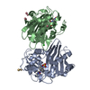
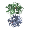
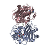
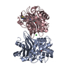
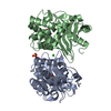
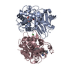
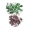
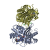
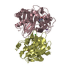
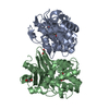
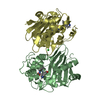
| Title | Crystal structure of OXA-48 beta-lactamase in the complex with the inhbitor ID2 | ||||||
 Components Components | Beta-lactamase | ||||||
 Keywords Keywords | HYDROLASE / Class D beta-lactamase / OXA-48 / antibiotic / dimer | ||||||
| Function / homology |  Function and homology information Function and homology informationpenicillin binding / antibiotic catabolic process / cell wall organization / beta-lactamase activity / beta-lactamase / response to antibiotic / plasma membrane Similarity search - Function | ||||||
| Biological species |  Klebsiella pneumoniae (bacteria) Klebsiella pneumoniae (bacteria) | ||||||
| Method |  X-RAY DIFFRACTION / SYNCHROTRON / MOLECULAR REPLACEMENT / Resolution: 2.05 Å X-RAY DIFFRACTION / SYNCHROTRON / MOLECULAR REPLACEMENT / Resolution: 2.05 Å | ||||||
 Authors Authors | Pochetti, G. / Montanari, R. / Capelli, D. / Garofalo, B. / Ombrato, R. | ||||||
 Citation Citation | Journal: Pharmaceuticals / Year: 2021 Title: Discovery of Novel Chemical Series of OXA-48 beta-Lactamase Inhibitors by High-Throughput Screening. Authors: Garofalo, B. / Prati, F. / Buonfiglio, R. / Coletta, I. / D'Atanasio, N. / Molteni, A. / Carettoni, D. / Wanke, V. / Pochetti, G. / Montanari, R. / Capelli, D. / Milanese, C. / Di Giorgio, F.P. / Ombrato, R. | ||||||
| History |
|
- Structure visualization
| Structure viewer | Molecule: MolmilJmol/JSmol |
|---|
- Downloads & links
-Download
| PDBx/mmCIF format | 7aux.cif.gz | 118.5 KB | Display | PDBx/mmCIF format |
|---|---|---|---|---|
| PDB format | pdb7aux.ent.gz | 90.7 KB | Display | PDB format |
| PDBx/mmJSON format | 7aux.json.gz | Tree view | PDBx/mmJSON format | |
| Others |  Other downloads Other downloads |
-Validation report
| Arichive directory | https://data.pdbj.org/pub/pdb/validation_reports/au/7auxftp://data.pdbj.org/pub/pdb/validation_reports/au/7aux | HTTPS FTP |
|---|
-Related structure data
| Related structure data |  7aw5C  3hbrS S: Starting model for refinement C: citing same article ( |
|---|---|
| Similar structure data |


-Links
 PDBj
PDBj- Assembly
| Deposited unit | 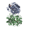
| ||||||||
|---|---|---|---|---|---|---|---|---|---|
| 1 |
| ||||||||
| Unit cell |
|
-Components
| #1: Protein | Mass: 28097.770 Da / Num. of mol.: 2 Source method: isolated from a genetically manipulated source Details: the Lysin 73 is carboxylated (KCX) / Source: (gene. exp.) Klebsiella pneumoniae (bacteria) / Gene: bla OXA-48, blaOXA-48, KPE71T_00045 / Production host: #2: Chemical | ChemComp-CL / |   Mass: 35.453 Da / Num. of mol.: 1 / Source method: obtained synthetically / Formula: Cl Mass: 35.453 Da / Num. of mol.: 1 / Source method: obtained synthetically / Formula: Cl#3: Chemical |   Mass: 387.388 Da / Num. of mol.: 2 / Source method: obtained synthetically / Formula: C22H17N3O4 Mass: 387.388 Da / Num. of mol.: 2 / Source method: obtained synthetically / Formula: C22H17N3O4#4: Water | ChemComp-HOH / |  Mass: 18.015 Da / Num. of mol.: 245 / Source method: isolated from a natural source / Formula: H2O Mass: 18.015 Da / Num. of mol.: 245 / Source method: isolated from a natural source / Formula: H2OHas ligand of interest | Y | |
|---|
-Experimental details
-Experiment
| Experiment | Method: X-RAY DIFFRACTION / Number of used crystals: 1 |
|---|
- Sample preparation
| Crystal | Density Matthews: 2.9 Å3/Da / Density % sol: 57.58 % |
|---|---|
| Crystal grow | Temperature: 293 K / Method: vapor diffusion, sitting drop / pH: 7.5 Details: 0.1 M Hepes, pH 7.5, 12% PEG8000, 8% 1-butanol, OXA-48 8.0-8.5 mg/ml, inhibitor/protein 6.5:1 |
-Data collection
| Diffraction | Mean temperature: 100 K / Serial crystal experiment: N |
|---|---|
| Diffraction source | Source: SYNCHROTRON / Site: ESRF  / Beamline: ID29 / Wavelength: 1.072 Å / Beamline: ID29 / Wavelength: 1.072 Å |
| Detector | Type: DECTRIS PILATUS3 S 6M / Detector: PIXEL / Date: Feb 18, 2016 |
| Radiation | Protocol: SINGLE WAVELENGTH / Monochromatic (M) / Laue (L): M / Scattering type: x-ray |
| Radiation wavelength | Wavelength: 1.072 Å / Relative weight: 1 |
| Reflection | Resolution: 2.05→62.882 Å / Num. obs: 40846 / % possible obs: 98.2 % / Redundancy: 3 % / Biso Wilson estimate: 27.9 Å2 / CC1/2: 0.994 / Rmerge(I) obs: 0.086 / Net I/σ(I): 8.2 |
| Reflection shell | Resolution: 2.05→2.11 Å / Rmerge(I) obs: 0.762 / Mean I/σ(I) obs: 1.8 / Num. unique obs: 2728 / CC1/2: 0.53 |
- Processing
| Software |
| ||||||||||||||||||||||||||||||||||||||||||||||||||||||||||||||||||||||||||||||||||||||||||
|---|---|---|---|---|---|---|---|---|---|---|---|---|---|---|---|---|---|---|---|---|---|---|---|---|---|---|---|---|---|---|---|---|---|---|---|---|---|---|---|---|---|---|---|---|---|---|---|---|---|---|---|---|---|---|---|---|---|---|---|---|---|---|---|---|---|---|---|---|---|---|---|---|---|---|---|---|---|---|---|---|---|---|---|---|---|---|---|---|---|---|---|
| Refinement | Method to determine structure: MOLECULAR REPLACEMENT Starting model: 3HBR Resolution: 2.05→62.882 Å / FOM work R set: 0.8009 / SU ML: 0.25 / Cross valid method: THROUGHOUT / σ(F): 1.34 / Phase error: 26.41 / Stereochemistry target values: ML
| ||||||||||||||||||||||||||||||||||||||||||||||||||||||||||||||||||||||||||||||||||||||||||
| Solvent computation | Shrinkage radii: 0.9 Å / VDW probe radii: 1.11 Å / Solvent model: FLAT BULK SOLVENT MODEL | ||||||||||||||||||||||||||||||||||||||||||||||||||||||||||||||||||||||||||||||||||||||||||
| Displacement parameters | Biso max: 78.01 Å2 / Biso mean: 34.92 Å2 / Biso min: 10.5 Å2 | ||||||||||||||||||||||||||||||||||||||||||||||||||||||||||||||||||||||||||||||||||||||||||
| Refinement step | Cycle: final / Resolution: 2.05→62.882 Å
| ||||||||||||||||||||||||||||||||||||||||||||||||||||||||||||||||||||||||||||||||||||||||||
| Refine LS restraints |
| ||||||||||||||||||||||||||||||||||||||||||||||||||||||||||||||||||||||||||||||||||||||||||
| LS refinement shell | Refine-ID: X-RAY DIFFRACTION / Rfactor Rfree error: 0
|
