[English] 日本語
 Yorodumi
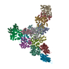
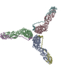
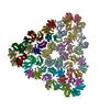
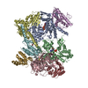
- PDB-7aqk: Model of the actin filament Arp2/3 complex branch junction in cells -
+ Open data
- Basic information
| Entry | Database: PDB / ID: 7aqk | ||||||
|---|---|---|---|---|---|---|---|
| Title | Model of the actin filament Arp2/3 complex branch junction in cells | ||||||
 Components Components |
| ||||||
 Keywords Keywords | STRUCTURAL PROTEIN / Actin / Arp2-3 complex / Cytoskeleton | ||||||
| Function / homology |  Function and homology information Function and homology informationpodosome core / apical tubulobulbar complex / basal ectoplasmic specialization / actin filament branch point / ventral surface of cell / microtubule organizing center localization / peripheral region of growth cone / negative regulation of bleb assembly / apical ectoplasmic specialization / regulation of myosin II filament organization ...podosome core / apical tubulobulbar complex / basal ectoplasmic specialization / actin filament branch point / ventral surface of cell / microtubule organizing center localization / peripheral region of growth cone / negative regulation of bleb assembly / apical ectoplasmic specialization / regulation of myosin II filament organization / tubulobulbar complex / concave side of sperm head / meiotic chromosome movement towards spindle pole / cytosolic transport / growth cone leading edge / lamellipodium organization / spindle localization / meiotic cytokinesis / positive regulation of barbed-end actin filament capping / protein kinase C signaling / leading edge of lamellipodium / postsynaptic actin cytoskeleton organization / EPHB-mediated forward signaling / hemidesmosome / muscle cell projection membrane / RHO GTPases Activate WASPs and WAVEs / actin filament network formation / asymmetric cell division / postsynapse organization / Arp2/3 protein complex / podosome ring / Arp2/3 complex-mediated actin nucleation / cellular response to rapamycin / Formation of the dystrophin-glycoprotein complex (DGC) / Striated Muscle Contraction / actin cap / negative regulation of axon extension / Regulation of actin dynamics for phagocytic cup formation / Clathrin-mediated endocytosis / maintenance of cell polarity / regulation of actin filament polymerization / positive regulation of astrocyte differentiation / positive regulation of dendritic spine morphogenesis / positive regulation of dendrite morphogenesis / apical dendrite / astrocyte differentiation / positive regulation of podosome assembly / positive regulation of synapse assembly / positive regulation of fibroblast migration / positive regulation of filopodium assembly / podosome / positive regulation of smooth muscle cell migration / smooth muscle cell migration / filamentous actin / mesenchyme migration / establishment or maintenance of cell polarity / positive regulation of actin filament polymerization / cell leading edge / brush border / striated muscle thin filament / skeletal muscle thin filament assembly / excitatory synapse / cilium assembly / glutamate receptor binding / positive regulation of protein targeting to membrane / regulation of synaptic vesicle endocytosis / cellular response to transforming growth factor beta stimulus / positive regulation of double-strand break repair via homologous recombination / positive regulation of lamellipodium assembly / skeletal muscle fiber development / stress fiber / cytoskeletal protein binding / cellular response to platelet-derived growth factor stimulus / ruffle / actin filament polymerization / Neutrophil degranulation / axon terminus / positive regulation of substrate adhesion-dependent cell spreading / positive regulation of neuron differentiation / secretory granule / cellular response to epidermal growth factor stimulus / dendritic shaft / regulation of actin cytoskeleton organization / positive regulation of protein localization to plasma membrane / meiotic cell cycle / cell projection / actin filament / filopodium / structural constituent of cytoskeleton / cellular response to type II interferon / Hydrolases; Acting on acid anhydrides; Acting on acid anhydrides to facilitate cellular and subcellular movement / Schaffer collateral - CA1 synapse / ruffle membrane / cell-cell junction / actin filament binding / cellular response to tumor necrosis factor / synaptic vesicle membrane / cell migration / lamellipodium / presynapse Similarity search - Function | ||||||
| Biological species |  | ||||||
| Method | ELECTRON MICROSCOPY / subtomogram averaging / cryo EM / Resolution: 9 Å | ||||||
 Authors Authors | Faessler, F. / Dimchev, G. / Hodirnau, V.V. / Wan, W. / Schur, F.K.M. | ||||||
| Funding support |  Austria, 1items Austria, 1items
| ||||||
 Citation Citation |  Journal: Nat Commun / Year: 2020 Journal: Nat Commun / Year: 2020Title: Cryo-electron tomography structure of Arp2/3 complex in cells reveals new insights into the branch junction. Authors: Florian Fäßler / Georgi Dimchev / Victor-Valentin Hodirnau / William Wan / Florian K M Schur /   Abstract: The actin-related protein (Arp)2/3 complex nucleates branched actin filament networks pivotal for cell migration, endocytosis and pathogen infection. Its activation is tightly regulated and involves ...The actin-related protein (Arp)2/3 complex nucleates branched actin filament networks pivotal for cell migration, endocytosis and pathogen infection. Its activation is tightly regulated and involves complex structural rearrangements and actin filament binding, which are yet to be understood. Here, we report a 9.0 Å resolution structure of the actin filament Arp2/3 complex branch junction in cells using cryo-electron tomography and subtomogram averaging. This allows us to generate an accurate model of the active Arp2/3 complex in the branch junction and its interaction with actin filaments. Notably, our model reveals a previously undescribed set of interactions of the Arp2/3 complex with the mother filament, significantly different to the previous branch junction model. Our structure also indicates a central role for the ArpC3 subunit in stabilizing the active conformation. | ||||||
| History |
|
- Structure visualization
| Movie |
 Movie viewer Movie viewer |
|---|---|
| Structure viewer | Molecule:  Molmil Molmil Jmol/JSmol Jmol/JSmol |
- Downloads & links
- Download
| PDBx/mmCIF format |  7aqk.cif.gz 7aqk.cif.gz | 790 KB | Display |  PDBx/mmCIF format PDBx/mmCIF format |
|---|---|---|---|---|
| PDB format |  pdb7aqk.ent.gz pdb7aqk.ent.gz | 505.9 KB | Display |  PDB format PDB format |
| PDBx/mmJSON format |  7aqk.json.gz 7aqk.json.gz | Tree view |  PDBx/mmJSON format PDBx/mmJSON format | |
| Others |  Other downloads Other downloads |
-Validation report
| Arichive directory |  https://data.pdbj.org/pub/pdb/validation_reports/aq/7aqk https://data.pdbj.org/pub/pdb/validation_reports/aq/7aqk ftp://data.pdbj.org/pub/pdb/validation_reports/aq/7aqk ftp://data.pdbj.org/pub/pdb/validation_reports/aq/7aqk | HTTPS FTP |
|---|
-Related structure data
| Related structure data |  11869MC C: citing same article ( M: map data used to model this data |
|---|---|
| Similar structure data |
- Links
Links
- Assembly
| Deposited unit | 
|
|---|---|
| 1 |
|
- Components
-Actin-related protein ... , 7 types, 7 molecules bacdefg
| #1: Protein | Mass: 44818.711 Da / Num. of mol.: 1 / Source method: isolated from a natural source Details: As models derived from Bos taurus Arp2/3 complexes have been used for fitting, also the Bos taurus sequence (as well as the corresponding UniProt identifier) is given here, even though they ...Details: As models derived from Bos taurus Arp2/3 complexes have been used for fitting, also the Bos taurus sequence (as well as the corresponding UniProt identifier) is given here, even though they were fit into a mouse structure. Source: (natural)  |
|---|---|
| #2: Protein | Mass: 47428.031 Da / Num. of mol.: 1 / Mutation: From Bos taurus to Mus Musculus I259V / Source method: isolated from a natural source Details: As models derived from Bos taurus Arp2/3 complexes have been used for fitting, also the Bos taurus sequence (as well as the corresponding UniProt identifier) is given here, even though they ...Details: As models derived from Bos taurus Arp2/3 complexes have been used for fitting, also the Bos taurus sequence (as well as the corresponding UniProt identifier) is given here, even though they were fit into a mouse structure. Source: (natural)  |
| #3: Protein | Mass: 41016.738 Da / Num. of mol.: 1 Mutation: From Bos taurus to Mus Musculus V43N, Q44K, V58I, D63E, K109N, S154N, N213S, A229V, S256N, S274N, G277V, K278T, L296M, S313G, A216T, R360K Source method: isolated from a natural source Details: As models derived from Bos taurus Arp2/3 complexes have been used for fitting, also the Bos taurus sequence (as well as the corresponding UniProt identifier) is given here, even though they ...Details: As models derived from Bos taurus Arp2/3 complexes have been used for fitting, also the Bos taurus sequence (as well as the corresponding UniProt identifier) is given here, even though they were fit into a mouse structure. Source: (natural)  |
| #4: Protein | Mass: 34402.043 Da / Num. of mol.: 1 / Mutation: From Bos taurus to Mus Musculus Y84F, S90P / Source method: isolated from a natural source Details: As models derived from Bos taurus Arp2/3 complexes have been used for fitting, also the Bos taurus sequence (as well as the corresponding UniProt identifier) is given here, even though they ...Details: As models derived from Bos taurus Arp2/3 complexes have been used for fitting, also the Bos taurus sequence (as well as the corresponding UniProt identifier) is given here, even though they were fit into a mouse structure. Source: (natural)  |
| #5: Protein | Mass: 20572.666 Da / Num. of mol.: 1 / Mutation: From Bos taurus to Mus Musculus I24L / Source method: isolated from a natural source Details: As models derived from Bos taurus Arp2/3 complexes have been used for fitting, also the Bos taurus sequence (as well as the corresponding UniProt identifier) is given here, even though they ...Details: As models derived from Bos taurus Arp2/3 complexes have been used for fitting, also the Bos taurus sequence (as well as the corresponding UniProt identifier) is given here, even though they were fit into a mouse structure. Source: (natural)  |
| #6: Protein | Mass: 19697.047 Da / Num. of mol.: 1 / Source method: isolated from a natural source Details: As models derived from Bos taurus Arp2/3 complexes have been used for fitting, also the Bos taurus sequence (as well as the corresponding UniProt identifier) is given here, even though they ...Details: As models derived from Bos taurus Arp2/3 complexes have been used for fitting, also the Bos taurus sequence (as well as the corresponding UniProt identifier) is given here, even though they were fit into a mouse structure. Source: (natural)  |
| #7: Protein | Mass: 16295.317 Da / Num. of mol.: 1 / Mutation: From Bos taurus to Mus Musculus D28E / Source method: isolated from a natural source Details: As models derived from Bos taurus Arp2/3 complexes have been used for fitting, also the Bos taurus sequence (as well as the corresponding UniProt identifier) is given here, even though they ...Details: As models derived from Bos taurus Arp2/3 complexes have been used for fitting, also the Bos taurus sequence (as well as the corresponding UniProt identifier) is given here, even though they were fit into a mouse structure. Source: (natural)  |
-Protein , 1 types, 11 molecules hijklmnopqr
| #8: Protein | Mass: 42096.953 Da / Num. of mol.: 11 / Source method: isolated from a natural source Details: As models derived from Oryctolagus cuniculus actin have been used for fitting, also the Oryctolagus cuniculus sequence (as well as the corresponding UniProt identifier) is given here, even ...Details: As models derived from Oryctolagus cuniculus actin have been used for fitting, also the Oryctolagus cuniculus sequence (as well as the corresponding UniProt identifier) is given here, even though they were fit into a mouse structure. Source: (natural)  |
|---|
-Experimental details
-Experiment
| Experiment | Method: ELECTRON MICROSCOPY |
|---|---|
| EM experiment | Aggregation state: CELL / 3D reconstruction method: subtomogram averaging |
- Sample preparation
| Component | Name: Actin Filament Arp2/3 Complex Branch Junction / Type: CELL Details: Structure obtained from the actin network of extracted and fixed mouse fibroblast lamellipodia Entity ID: all / Source: NATURAL | ||||||||||||||||||||||||
|---|---|---|---|---|---|---|---|---|---|---|---|---|---|---|---|---|---|---|---|---|---|---|---|---|---|
| Source (natural) | Organism:  | ||||||||||||||||||||||||
| Buffer solution | pH: 6.1 / Details: Adjust to pH 6.1 using NaOH | ||||||||||||||||||||||||
| Buffer component |
| ||||||||||||||||||||||||
| Specimen | Embedding applied: NO / Shadowing applied: NO / Staining applied: NO / Vitrification applied: YES | ||||||||||||||||||||||||
| Specimen support | Details: After glow discharging of the grid and prior to the seeding of cells, the grid was coated using 25ug/ml Fibronectin Grid material: GOLD / Grid mesh size: 200 divisions/in. / Grid type: Quantifoil R2/2 | ||||||||||||||||||||||||
| Vitrification | Cryogen name: ETHANE / Humidity: 80 % / Chamber temperature: 277 K Details: Leica GP2, 3,5sec back-blotting, sensor on, 0,1mm movement after contact, manually pre-blotted within the chamber prior to the application of fiducials |
- Electron microscopy imaging
| Experimental equipment |  Model: Titan Krios / Image courtesy: FEI Company |
|---|---|
| Microscopy | Model: FEI TITAN KRIOS |
| Electron gun | Electron source:  FIELD EMISSION GUN / Accelerating voltage: 300 kV / Illumination mode: FLOOD BEAM FIELD EMISSION GUN / Accelerating voltage: 300 kV / Illumination mode: FLOOD BEAM |
| Electron lens | Mode: BRIGHT FIELD / Nominal magnification: 42000 X / Nominal defocus max: -5.5 nm / Nominal defocus min: -1.75 nm / Cs: 2.7 mm / C2 aperture diameter: 50 µm / Alignment procedure: ZEMLIN TABLEAU |
| Specimen holder | Cryogen: NITROGEN / Specimen holder model: FEI TITAN KRIOS AUTOGRID HOLDER |
| Image recording | Average exposure time: 1.21 sec. / Electron dose: 2.79 e/Å2 / Film or detector model: GATAN K3 BIOQUANTUM (6k x 4k) Details: Images were collected in movie-mode at 7 frames per tilt |
| EM imaging optics | Energyfilter name: GIF Bioquantum / Energyfilter slit width: 20 eV |
- Processing
| EM software |
| ||||||||||||||||||||||||||||||||||||||||||||||||||||||||||||||||||||||||||||||||||||||||||||||||||||||||||||||||||||||||||||||
|---|---|---|---|---|---|---|---|---|---|---|---|---|---|---|---|---|---|---|---|---|---|---|---|---|---|---|---|---|---|---|---|---|---|---|---|---|---|---|---|---|---|---|---|---|---|---|---|---|---|---|---|---|---|---|---|---|---|---|---|---|---|---|---|---|---|---|---|---|---|---|---|---|---|---|---|---|---|---|---|---|---|---|---|---|---|---|---|---|---|---|---|---|---|---|---|---|---|---|---|---|---|---|---|---|---|---|---|---|---|---|---|---|---|---|---|---|---|---|---|---|---|---|---|---|---|---|---|
| CTF correction | Type: PHASE FLIPPING AND AMPLITUDE CORRECTION | ||||||||||||||||||||||||||||||||||||||||||||||||||||||||||||||||||||||||||||||||||||||||||||||||||||||||||||||||||||||||||||||
| Symmetry | Point symmetry: C1 (asymmetric) | ||||||||||||||||||||||||||||||||||||||||||||||||||||||||||||||||||||||||||||||||||||||||||||||||||||||||||||||||||||||||||||||
| 3D reconstruction | Resolution: 9 Å / Resolution method: FSC 0.143 CUT-OFF / Num. of particles: 14296 Details: Final reconstruction in RELION was performed after Multiple particle refinement in M version 1.0.9. Num. of class averages: 1 / Symmetry type: POINT | ||||||||||||||||||||||||||||||||||||||||||||||||||||||||||||||||||||||||||||||||||||||||||||||||||||||||||||||||||||||||||||||
| EM volume selection | Method: Template Matching Details: After first classification in Dynamo and re-extraction in Warp 17,146 subvolumes remained. Num. of tomograms: 131 / Num. of volumes extracted: 39300 Reference model: Reference generated from manually selected particles | ||||||||||||||||||||||||||||||||||||||||||||||||||||||||||||||||||||||||||||||||||||||||||||||||||||||||||||||||||||||||||||||
| Atomic model building | Protocol: FLEXIBLE FIT / Space: REAL | ||||||||||||||||||||||||||||||||||||||||||||||||||||||||||||||||||||||||||||||||||||||||||||||||||||||||||||||||||||||||||||||
| Atomic model building | 3D fitting-ID: 1 / Source name: PDB / Type: experimental model
|
 Movie
Movie Controller


 PDBj
PDBj


