 Movie
Movie Controller


+ Open data

- Basic information
| Entry | Database: PDB / ID: 7ap7 | ||||||
|---|---|---|---|---|---|---|---|
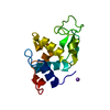
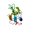
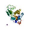
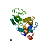
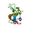
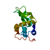
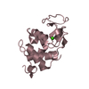
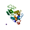
| Title | Structure of the W64R amyloidogenic variant of human lysozyme | ||||||
 Components Components | Lysozyme C | ||||||
 Keywords Keywords | HYDROLASE / lysozyme / amyloidoses | ||||||
| Function / homology |  Function and homology information Function and homology informationantimicrobial humoral response / Antimicrobial peptides / metabolic process / specific granule lumen / azurophil granule lumen / tertiary granule lumen / lysozyme / lysozyme activity / defense response to Gram-negative bacterium / killing of cells of another organism ...antimicrobial humoral response / Antimicrobial peptides / metabolic process / specific granule lumen / azurophil granule lumen / tertiary granule lumen / lysozyme / lysozyme activity / defense response to Gram-negative bacterium / killing of cells of another organism / defense response to Gram-positive bacterium / defense response to bacterium / inflammatory response / Amyloid fiber formation / Neutrophil degranulation / extracellular space / extracellular exosome / extracellular region / identical protein binding Similarity search - Function | ||||||
| Biological species |  Homo sapiens (human) Homo sapiens (human) | ||||||
| Method |  X-RAY DIFFRACTION / SYNCHROTRON / MOLECULAR REPLACEMENT / molecular replacement / Resolution: 1.15 Å X-RAY DIFFRACTION / SYNCHROTRON / MOLECULAR REPLACEMENT / molecular replacement / Resolution: 1.15 Å | ||||||
 Authors Authors | Vettore, N. / Herman, R. / Kerff, F. / Charlier, P. / Sauvage, E. / Brans, A. / Morray, J. / Dobson, C. / Kumita, J. / Dumoulin, M. | ||||||
 Citation Citation | Journal: Biophys.Chem. / Year: 2021 Title: Characterisation of the structural, dynamic and aggregation properties of the W64R amyloidogenic variant of human lysozyme. Authors: Vettore, N. / Moray, J. / Brans, A. / Herman, R. / Charlier, P. / Kumita, J.R. / Kerff, F. / Dobson, C.M. / Dumoulin, M. | ||||||
| History |
|
- Structure visualization
| Structure viewer | Molecule: MolmilJmol/JSmol |
|---|
- Downloads & links
-Download
| PDBx/mmCIF format | 7ap7.cif.gz | 98.7 KB | Display | PDBx/mmCIF format |
|---|---|---|---|---|
| PDB format | pdb7ap7.ent.gz | 76 KB | Display | PDB format |
| PDBx/mmJSON format | 7ap7.json.gz | Tree view | PDBx/mmJSON format | |
| Others |  Other downloads Other downloads |
-Validation report
| Summary document | 7ap7_validation.pdf.gz | 316.2 KB | Display | wwPDB validaton report |
|---|---|---|---|---|
| Full document | 7ap7_full_validation.pdf.gz | 316.2 KB | Display | |
| Data in XML | 7ap7_validation.xml.gz | 9.3 KB | Display | |
| Data in CIF | 7ap7_validation.cif.gz | 13.2 KB | Display | |
| Arichive directory | https://data.pdbj.org/pub/pdb/validation_reports/ap/7ap7ftp://data.pdbj.org/pub/pdb/validation_reports/ap/7ap7 | HTTPS FTP |
-Related structure data
| Related structure data | 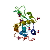 1jsfS S: Starting model for refinement |
|---|---|
| Similar structure data |


-Links
 PDBj
PDBj


- Assembly
| Deposited unit | 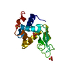
| ||||||||
|---|---|---|---|---|---|---|---|---|---|
| 1 |
| ||||||||
| Unit cell |
|
-Components
| #1: Protein | Mass: 14691.678 Da / Num. of mol.: 1 / Mutation: W64R Source method: isolated from a genetically manipulated source Source: (gene. exp.) Homo sapiens (human) / Gene: LYZ, LZM / Production host:  Komagataella pastoris (fungus) / References: UniProt: P61626, lysozyme Komagataella pastoris (fungus) / References: UniProt: P61626, lysozyme | ||||
|---|---|---|---|---|---|
| #2: Chemical |   Mass: 96.063 Da / Num. of mol.: 3 / Source method: obtained synthetically / Formula: SO4 Mass: 96.063 Da / Num. of mol.: 3 / Source method: obtained synthetically / Formula: SO4#3: Water | ChemComp-HOH / |  Mass: 18.015 Da / Num. of mol.: 164 / Source method: isolated from a natural source / Formula: H2O Mass: 18.015 Da / Num. of mol.: 164 / Source method: isolated from a natural source / Formula: H2OHas ligand of interest | N | |
-Experimental details
-Experiment
| Experiment | Method: X-RAY DIFFRACTION / Number of used crystals: 1 |
|---|
- Sample preparation
| Crystal | Density Matthews: 1.86 Å3/Da / Density % sol: 33.73 % |
|---|---|
| Crystal grow | Temperature: 293 K / Method: vapor diffusion, sitting drop / pH: 6.5 Details: 1.25M (NH4)2SO4, 0.1M cacodylate pH 6.5, 10 mM Tris pH 7, 14.4 TP buffer |
-Data collection
| Diffraction | Mean temperature: 100 K / Serial crystal experiment: N | ||||||||||||||||||||||||||||||||||||||||||||||||||||||||||||||||||||||
|---|---|---|---|---|---|---|---|---|---|---|---|---|---|---|---|---|---|---|---|---|---|---|---|---|---|---|---|---|---|---|---|---|---|---|---|---|---|---|---|---|---|---|---|---|---|---|---|---|---|---|---|---|---|---|---|---|---|---|---|---|---|---|---|---|---|---|---|---|---|---|---|
| Diffraction source | Source: SYNCHROTRON / Site: SOLEIL  / Beamline: PROXIMA 2 / Wavelength: 0.980119943619 Å / Beamline: PROXIMA 2 / Wavelength: 0.980119943619 Å | ||||||||||||||||||||||||||||||||||||||||||||||||||||||||||||||||||||||
| Detector | Type: DECTRIS EIGER X 9M / Detector: PIXEL / Date: Jun 10, 2016 | ||||||||||||||||||||||||||||||||||||||||||||||||||||||||||||||||||||||
| Radiation | Protocol: SINGLE WAVELENGTH / Monochromatic (M) / Laue (L): M / Scattering type: x-ray | ||||||||||||||||||||||||||||||||||||||||||||||||||||||||||||||||||||||
| Radiation wavelength | Wavelength: 0.980119943619 Å / Relative weight: 1 | ||||||||||||||||||||||||||||||||||||||||||||||||||||||||||||||||||||||
| Reflection | Resolution: 1.15→41.384 Å / Num. obs: 39372 / % possible obs: 98.7 % / Observed criterion σ(I): -3 / Redundancy: 6.35 % / Biso Wilson estimate: 12.31 Å2 / CC1/2: 0.998 / Rmerge(I) obs: 0.084 / Net I/σ(I): 10.04 | ||||||||||||||||||||||||||||||||||||||||||||||||||||||||||||||||||||||
| Reflection shell |
|
-Phasing
| Phasing | Method: molecular replacement | |||||||||
|---|---|---|---|---|---|---|---|---|---|---|
| Phasing MR |
|
- Processing
| Software |
| |||||||||||||||||||||||||||||||||||||||||||||||||||||||||||||||||||||||||||||||||||||||||||||||||||||||||
|---|---|---|---|---|---|---|---|---|---|---|---|---|---|---|---|---|---|---|---|---|---|---|---|---|---|---|---|---|---|---|---|---|---|---|---|---|---|---|---|---|---|---|---|---|---|---|---|---|---|---|---|---|---|---|---|---|---|---|---|---|---|---|---|---|---|---|---|---|---|---|---|---|---|---|---|---|---|---|---|---|---|---|---|---|---|---|---|---|---|---|---|---|---|---|---|---|---|---|---|---|---|---|---|---|---|---|
| Refinement | Method to determine structure: MOLECULAR REPLACEMENT Starting model: 1JSF Resolution: 1.15→30.298 Å / SU ML: 0.17 / Cross valid method: FREE R-VALUE / σ(F): 1.38 / Phase error: 19.07
| |||||||||||||||||||||||||||||||||||||||||||||||||||||||||||||||||||||||||||||||||||||||||||||||||||||||||
| Solvent computation | Shrinkage radii: 0.47 Å / VDW probe radii: 0.8 Å / Bsol: 45.093 Å2 / ksol: 0.439 e/Å3 | |||||||||||||||||||||||||||||||||||||||||||||||||||||||||||||||||||||||||||||||||||||||||||||||||||||||||
| Displacement parameters | Biso max: 47.65 Å2 / Biso mean: 16.58 Å2 / Biso min: 6.28 Å2
| |||||||||||||||||||||||||||||||||||||||||||||||||||||||||||||||||||||||||||||||||||||||||||||||||||||||||
| Refinement step | Cycle: final / Resolution: 1.15→30.298 Å
| |||||||||||||||||||||||||||||||||||||||||||||||||||||||||||||||||||||||||||||||||||||||||||||||||||||||||
| Refine LS restraints |
| |||||||||||||||||||||||||||||||||||||||||||||||||||||||||||||||||||||||||||||||||||||||||||||||||||||||||
| LS refinement shell |
|
