 Movie
Movie Controller


+ Open data

- Basic information
| Entry | Database: PDB / ID: 1i22 | ||||||
|---|---|---|---|---|---|---|---|
| Title | MUTANT HUMAN LYSOZYME (A83K/Q86D/A92D) | ||||||
 Components Components | LYSOZYME C | ||||||
 Keywords Keywords | HYDROLASE / CALCIUM BINDING SITE / MUTANT HUMAN LYSOZYME | ||||||
| Function / homology |  Function and homology information Function and homology informationantimicrobial humoral response / Antimicrobial peptides / specific granule lumen / lysozyme / lysozyme activity / azurophil granule lumen / tertiary granule lumen / killing of cells of another organism / defense response to Gram-negative bacterium / defense response to bacterium ...antimicrobial humoral response / Antimicrobial peptides / specific granule lumen / lysozyme / lysozyme activity / azurophil granule lumen / tertiary granule lumen / killing of cells of another organism / defense response to Gram-negative bacterium / defense response to bacterium / defense response to Gram-positive bacterium / inflammatory response / Amyloid fiber formation / Neutrophil degranulation / : / extracellular exosome / extracellular region / identical protein binding Similarity search - Function | ||||||
| Biological species |  Homo sapiens (human) Homo sapiens (human) | ||||||
| Method |  X-RAY DIFFRACTION / MOLECULAR REPLACEMENT / Resolution: 1.8 Å X-RAY DIFFRACTION / MOLECULAR REPLACEMENT / Resolution: 1.8 Å | ||||||
 Authors Authors | Kuroki, R. | ||||||
 Citation Citation | Journal: J.Biol.Chem. / Year: 1998 Title: Structural and thermodynamic responses of mutations at a Ca2+ binding site engineered into human lysozyme. Authors: Kuroki, R. / Yutani, K. #1: Journal: J.Biol.Chem. / Year: 1992Title: Thermodynamic changes in the binding of Ca2+ to a mutant human lysozyme (D86/92). Enthalpy-entropy compensation observed upon Ca2+ binding to proteins. Authors: Kuroki, R. / Nitta, K. / Yutani, K. #2: Journal: Proc.Natl.Acad.Sci.USA / Year: 1992Title: Entropic Stabilization of a Mutant Human Lysozyme Induced by Calcium Binding Authors: Kuroki, R. / Kawakita, S. / Nakamura, H. / Yutani, K. #3: Journal: J.Biol.Chem. / Year: 1991Title: Crystal Structures of the Apo- and Holomutant Human Lysozymes with an Introduced Ca2+ Binding Site Authors: Inaka, K. / Kuroki, R. / Kikuchi, M. / Matsushima, M. #4: Journal: Proc.Natl.Acad.Sci.USA / Year: 1989Title: Design and Creation of a Ca2+ Binding Site in Human Lysozyme to Enhanced Structural Stability Authors: Kuroki, R. / Taniyama, Y. / Seko, C. / Nakamura, H. / Kikuchi, M. / Ikehara, M. | ||||||
| History |
|
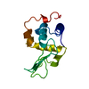
- Structure visualization
| Structure viewer | Molecule: MolmilJmol/JSmol |
|---|
- Downloads & links
-Download
| PDBx/mmCIF format | 1i22.cif.gz | 122.4 KB | Display | PDBx/mmCIF format |
|---|---|---|---|---|
| PDB format | pdb1i22.ent.gz | 94.6 KB | Display | PDB format |
| PDBx/mmJSON format | 1i22.json.gz | Tree view | PDBx/mmJSON format | |
| Others |  Other downloads Other downloads |
-Validation report
| Arichive directory | https://data.pdbj.org/pub/pdb/validation_reports/i2/1i22ftp://data.pdbj.org/pub/pdb/validation_reports/i2/1i22 | HTTPS FTP |
|---|
-Related structure data
| Related structure data |  1i1zC  1i20C  3lhmS C: citing same article ( S: Starting model for refinement |
|---|---|
| Similar structure data |


-Links
 PDBj
PDBj

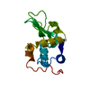
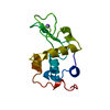
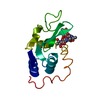
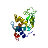
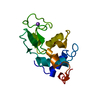
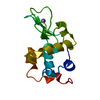
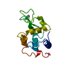
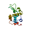
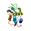
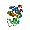
- Assembly
| Deposited unit | 
| ||||||||
|---|---|---|---|---|---|---|---|---|---|
| 1 | 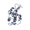
| ||||||||
| 2 | 
| ||||||||
| 3 | 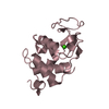
| ||||||||
| 4 | 
| ||||||||
| Unit cell |
|
-Components
| #1: Protein | Mass: 14809.764 Da / Num. of mol.: 4 / Mutation: A83K/Q86D/A92D Source method: isolated from a genetically manipulated source Source: (gene. exp.) Homo sapiens (human) / Plasmid: PERI8811 / Production host:  #2: Chemical | ChemComp-CA /   Mass: 40.078 Da / Num. of mol.: 4 / Source method: obtained synthetically / Formula: Ca Mass: 40.078 Da / Num. of mol.: 4 / Source method: obtained synthetically / Formula: Ca#3: Water | ChemComp-HOH / |  Mass: 18.015 Da / Num. of mol.: 317 / Source method: isolated from a natural source / Formula: H2O Mass: 18.015 Da / Num. of mol.: 317 / Source method: isolated from a natural source / Formula: H2OHas protein modification | Y | |
|---|
-Experimental details
-Experiment
| Experiment | Method: X-RAY DIFFRACTION / Number of used crystals: 1 |
|---|
- Sample preparation
| Crystal | Density Matthews: 2.35 Å3/Da / Density % sol: 47.63 % | |||||||||||||||||||||||||||||||||||
|---|---|---|---|---|---|---|---|---|---|---|---|---|---|---|---|---|---|---|---|---|---|---|---|---|---|---|---|---|---|---|---|---|---|---|---|---|
| Crystal grow | Temperature: 298 K / Method: vapor diffusion, hanging drop / pH: 6 Details: PHOSPHATE, SODIUM CHLORIDE, CALCIUM CHLORIDE, pH 6.00, VAPOR DIFFUSION, HANGING DROP, temperature 298K | |||||||||||||||||||||||||||||||||||
| Crystal grow | *PLUS Method: unknown / pH: 6 | |||||||||||||||||||||||||||||||||||
| Components of the solutions | *PLUS
|
-Data collection
| Diffraction | Mean temperature: 293 K |
|---|---|
| Diffraction source | Source: ROTATING ANODE / Type: RIGAKU RU200 / Wavelength: 1.5418 / Wavelength: 1.5418 Å |
| Detector | Type: SDMS / Detector: AREA DETECTOR / Date: Feb 26, 1993 |
| Radiation | Protocol: SINGLE WAVELENGTH / Monochromatic (M) / Laue (L): M / Scattering type: x-ray |
| Radiation wavelength | Wavelength: 1.5418 Å / Relative weight: 1 |
| Reflection | Resolution: 1.8→50 Å / Num. obs: 39085 / Rmerge(I) obs: 0.041 |
| Reflection | *PLUS Highest resolution: 1.8 Å / Lowest resolution: 9999 Å / Num. measured all: 66509 |
- Processing
| Software |
| ||||||||||||||||||||||||||||||||||||||||||||||||||
|---|---|---|---|---|---|---|---|---|---|---|---|---|---|---|---|---|---|---|---|---|---|---|---|---|---|---|---|---|---|---|---|---|---|---|---|---|---|---|---|---|---|---|---|---|---|---|---|---|---|---|---|
| Refinement | Method to determine structure: MOLECULAR REPLACEMENT Starting model: 3LHM (HOLO-Q86D/A92D MUTANT LYSOZYME) Resolution: 1.8→20 Å / σ(F): 0
| ||||||||||||||||||||||||||||||||||||||||||||||||||
| Refinement step | Cycle: LAST / Resolution: 1.8→20 Å
| ||||||||||||||||||||||||||||||||||||||||||||||||||
| Refine LS restraints |
| ||||||||||||||||||||||||||||||||||||||||||||||||||
| Software | *PLUS Name: TNT / Classification: refinement | ||||||||||||||||||||||||||||||||||||||||||||||||||
| Refinement | *PLUS Lowest resolution: 20 Å / σ(F): 0 / Rfactor Rwork: 0.172 | ||||||||||||||||||||||||||||||||||||||||||||||||||
| Solvent computation | *PLUS | ||||||||||||||||||||||||||||||||||||||||||||||||||
| Displacement parameters | *PLUS | ||||||||||||||||||||||||||||||||||||||||||||||||||
| Refine LS restraints | *PLUS
|
