 ムービー
ムービー コントローラー


+ データを開く

- 基本情報
基本情報
| 登録情報 | データベース: PDB / ID: 7aoy | ||||||
|---|---|---|---|---|---|---|---|
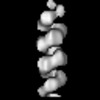
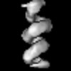
| タイトル | Helical arrangement of Bunyamwera virus nucleocapsid protein within a native ribonucleoprotein | ||||||
 要素 要素 | Nucleoprotein | ||||||
 キーワード キーワード | VIRAL PROTEIN / helix / ribonucleoprotein / nucleocapsid / virus | ||||||
| 機能・相同性 |  機能・相同性情報 機能・相同性情報helical viral capsid / viral nucleocapsid / ribonucleoprotein complex / symbiont-mediated suppression of host gene expression / RNA binding 類似検索 - 分子機能 | ||||||
| 生物種 |  Bunyamwera virus (ブニヤンベラウイルス) Bunyamwera virus (ブニヤンベラウイルス) | ||||||
| 手法 | 電子顕微鏡法 / 単粒子再構成法 / クライオ電子顕微鏡法 / 解像度: 13 Å | ||||||
 データ登録者 データ登録者 | Hopkins, F.R. / Fontana, J. / Barr, J.N. | ||||||
| 資金援助 |  英国, 1件 英国, 1件
| ||||||
 引用 引用 | ジャーナル: mBio / 年: 2022 タイトル: The Native Orthobunyavirus Ribonucleoprotein Possesses a Helical Architecture. 著者: Francis R Hopkins / Beatriz Álvarez-Rodríguez / George R Heath / Kyriakoulla Panayi / Samantha Hover / Thomas A Edwards / John N Barr / Juan Fontana / 要旨: The order is the largest group of negative-sense RNA viruses, containing many lethal human pathogens for which approved anti-infective measures are not available. The bunyavirus genome consists of ...The order is the largest group of negative-sense RNA viruses, containing many lethal human pathogens for which approved anti-infective measures are not available. The bunyavirus genome consists of multiple negative-sense RNA segments enwrapped by the virus-encoded nucleocapsid protein (NP), which together with the viral polymerase form ribonucleoproteins (RNPs). RNPs represent substrates for RNA synthesis and virion assembly, which require inherent flexibility, consistent with the appearance of RNPs spilled from virions. These observations have resulted in conflicting models describing the overall RNP architecture. Here, we purified RNPs from Bunyamwera virus (BUNV), the prototypical orthobunyavirus. The lengths of purified RNPs imaged by negative staining resulted in 3 populations of RNPs, suggesting that RNPs possess a consistent method of condensation. Employing microscopy approaches, we conclusively show that the NP portion of BUNV RNPs is helical. Furthermore, we present a pseudo-atomic model for this portion based on a cryo-electron microscopy average at 13 Å resolution, which allowed us to fit the BUNV NP crystal structure by molecular dynamics. This model was confirmed by NP mutagenesis using a mini-genome system. The model shows that adjacent NP monomers in the RNP chain interact laterally through flexible N- and C-terminal arms only, with no longitudinal helix-stabilizing interactions, thus providing a potential model for the molecular basis for RNP flexibility. Excessive RNase treatment disrupts native RNPs, suggesting that RNA was key in maintaining the RNP structure. Overall, this work will inform studies on bunyaviral RNP assembly, packaging, and RNA replication, and aid in future antiviral strategies. Bunyaviruses are emerging RNA viruses that cause significant disease and economic burden and for which vaccines or therapies approved for humans are not available. The bunyavirus genome is wrapped up by the nucleoprotein (NP) and interacts with the viral polymerase, forming a ribonucleoprotein (RNP). This is the only form of the genome active for viral replication and assembly. However, until now how NPs are organized within an RNP was not known for any orthobunyavirus. Here, we purified RNPs from the prototypical orthobunyavirus, Bunyamwera virus, and employed microscopy approaches to show that the NP portion of the RNP was helical. We then combined our helical average with the known structure of an NP monomer, generating a pseudo-atomic model of this region. This arrangement allowed the RNPs to be highly flexible, which was critical for several stages of the viral replication cycle, such as segment circularization. | ||||||
| 履歴 |
|
- 構造の表示
構造の表示
| 構造ビューア | 分子: MolmilJmol/JSmol |
|---|
- ダウンロードとリンク
-ダウンロード
| PDBx/mmCIF形式 | 7aoy.cif.gz | 354 KB | 表示 | PDBx/mmCIF形式 |
|---|---|---|---|---|
| PDB形式 | pdb7aoy.ent.gz | 282 KB | 表示 | PDB形式 |
| PDBx/mmJSON形式 | 7aoy.json.gz | ツリー表示 | PDBx/mmJSON形式 | |
| その他 |  その他のダウンロード その他のダウンロード |
-検証レポート
| アーカイブディレクトリ | https://data.pdbj.org/pub/pdb/validation_reports/ao/7aoyftp://data.pdbj.org/pub/pdb/validation_reports/ao/7aoy | HTTPS FTP |
|---|
-関連構造データ
-リンク
 PDBj
PDBj- 集合体
集合体
| 登録構造単位 | 
|
|---|---|
| 1 |
|
-要素
| #1: タンパク質 | 分子量: 26698.760 Da / 分子数: 9 / 由来タイプ: 天然 由来: (天然) Bunyamwera virus (ブニヤンベラウイルス)細胞株: BHK-21 / 参照: UniProt: P16495 |
|---|
-実験情報
-実験
| 実験 | 手法: 電子顕微鏡法 |
|---|---|
| EM実験 | 試料の集合状態: FILAMENT / 3次元再構成法: 単粒子再構成法 |
- 試料調製
試料調製
| 構成要素 | 名称: Native ribonucleoprotein extracted from Bunyamwera virus タイプ: COMPLEX / Entity ID: all / 由来: NATURAL |
|---|---|
| 分子量 | 実験値: NO |
| 由来(天然) | 生物種: Bunyamwera virus (ブニヤンベラウイルス) |
| 緩衝液 | pH: 7.4 |
| 試料 | 包埋: NO / シャドウイング: NO / 染色: NO / 凍結: YES |
| 急速凍結 | 装置: FEI VITROBOT MARK II / 凍結剤: NITROGEN |
- 電子顕微鏡撮影
| 実験機器 |  モデル: Titan Krios / 画像提供: FEI Company |
|---|---|
| 顕微鏡 | モデル: FEI TITAN KRIOS |
| 電子銃 | 電子線源:  FIELD EMISSION GUN / 加速電圧: 300 kV / 照射モード: FLOOD BEAM FIELD EMISSION GUN / 加速電圧: 300 kV / 照射モード: FLOOD BEAM |
| 電子レンズ | モード: BRIGHT FIELD / 倍率(公称値): 130000 X |
| 撮影 | 平均露光時間: 9 sec. / 電子線照射量: 51.3 e/Å2 / 検出モード: COUNTING フィルム・検出器のモデル: GATAN K2 SUMMIT (4k x 4k) |
| 電子光学装置 | 位相板: VOLTA PHASE PLATE |
- 解析
解析
| EMソフトウェア |
| ||||||||||||
|---|---|---|---|---|---|---|---|---|---|---|---|---|---|
| CTF補正 | タイプ: PHASE FLIPPING AND AMPLITUDE CORRECTION | ||||||||||||
| 3次元再構成 | 解像度: 13 Å / 解像度の算出法: FSC 0.143 CUT-OFF / 粒子像の数: 5077 / 対称性のタイプ: POINT | ||||||||||||
| 原子モデル構築 | プロトコル: FLEXIBLE FIT | ||||||||||||
| 原子モデル構築 | PDB-ID: 3ZLA PDB chain-ID: B / Accession code: 3ZLA / Source name: PDB / タイプ: experimental model |

