 Movie
Movie Controller

[English] 日本語
 Yorodumi
- PDB-7ao2: Crystal structure of CotB2 variant W288G in complex with alendronate -
+ Open data

- Basic information
| Entry | Database: PDB / ID: 7ao2 | ||||||
|---|---|---|---|---|---|---|---|
| Title | Crystal structure of CotB2 variant W288G in complex with alendronate | ||||||
 Components Components | Cyclooctat-9-en-7-ol synthase | ||||||
 Keywords Keywords | LYASE / TERPENE SYNTHASE / CYCLOOCTATIN | ||||||
| Function / homology |  Function and homology information Function and homology informationcyclooctat-9-en-7-ol synthase / isomerase activity / lyase activity / metal ion binding Similarity search - Function | ||||||
| Biological species |  Streptomyces melanosporofaciens (bacteria) Streptomyces melanosporofaciens (bacteria) | ||||||
| Method |  X-RAY DIFFRACTION / SYNCHROTRON / MOLECULAR REPLACEMENT / Resolution: 1.75 Å X-RAY DIFFRACTION / SYNCHROTRON / MOLECULAR REPLACEMENT / Resolution: 1.75 Å | ||||||
 Authors Authors | Dimos, N. / Driller, R. / Loll, B. | ||||||
| Funding support |  Germany, 1items Germany, 1items
| ||||||
 Citation Citation | Journal: J.Am.Chem.Soc. / Year: 2020 Title: The Impression of a Nonexisting Catalytic Effect: The Role of CotB2 in Guiding the Complex Biosynthesis of Cyclooctat-9-en-7-ol. Authors: Raz, K. / Driller, R. / Dimos, N. / Ringel, M. / Bruck, T. / Loll, B. / Major, D.T. | ||||||
| History |
|
- Structure visualization
| Structure viewer | Molecule: MolmilJmol/JSmol |
|---|
- Downloads & links
-Download
| PDBx/mmCIF format | 7ao2.cif.gz | 176.4 KB | Display | PDBx/mmCIF format |
|---|---|---|---|---|
| PDB format | pdb7ao2.ent.gz | 111.5 KB | Display | PDB format |
| PDBx/mmJSON format | 7ao2.json.gz | Tree view | PDBx/mmJSON format | |
| Others |  Other downloads Other downloads |
-Validation report
| Arichive directory | https://data.pdbj.org/pub/pdb/validation_reports/ao/7ao2ftp://data.pdbj.org/pub/pdb/validation_reports/ao/7ao2 | HTTPS FTP |
|---|
-Related structure data
| Related structure data | 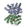 7ao0C 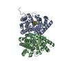 7ao1C 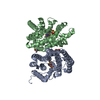 7ao3C 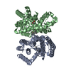 7ao4C  7ao5C  6ggjS S: Starting model for refinement C: citing same article ( |
|---|---|
| Similar structure data |


-Links
 PDBj
PDBj
- Assembly
| Deposited unit | 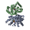
| ||||||||||||
|---|---|---|---|---|---|---|---|---|---|---|---|---|---|
| 1 |
| ||||||||||||
| Unit cell |
|
-Components
-Protein , 1 types, 2 molecules AB
| #1: Protein | Mass: 36641.363 Da / Num. of mol.: 2 / Mutation: W288G Source method: isolated from a genetically manipulated source Source: (gene. exp.) Streptomyces melanosporofaciens (bacteria)Gene: CotB2 / Production host: |
|---|
-Non-polymers , 5 types, 408 molecules 


| #2: Chemical |  Mass: 118.174 Da / Num. of mol.: 2 / Source method: obtained synthetically / Formula: C6H14O2 / Comment: precipitant*YM Mass: 118.174 Da / Num. of mol.: 2 / Source method: obtained synthetically / Formula: C6H14O2 / Comment: precipitant*YM#3: Chemical |  Mass: 245.064 Da / Num. of mol.: 2 / Source method: obtained synthetically / Formula: C4H9NO7P2 / Comment: medication*YM Mass: 245.064 Da / Num. of mol.: 2 / Source method: obtained synthetically / Formula: C4H9NO7P2 / Comment: medication*YM#4: Chemical | ChemComp-MG /  Mass: 24.305 Da / Num. of mol.: 6 / Source method: obtained synthetically / Formula: Mg Mass: 24.305 Da / Num. of mol.: 6 / Source method: obtained synthetically / Formula: Mg#5: Chemical |  Mass: 35.453 Da / Num. of mol.: 2 / Source method: obtained synthetically / Formula: Cl Mass: 35.453 Da / Num. of mol.: 2 / Source method: obtained synthetically / Formula: Cl#6: Water | ChemComp-HOH / | Mass: 18.015 Da / Num. of mol.: 396 / Source method: isolated from a natural source / Formula: H2O |
|---|
-Details
| Has ligand of interest | N |
|---|
-Experimental details
-Experiment
| Experiment | Method: X-RAY DIFFRACTION / Number of used crystals: 1 |
|---|
- Sample preparation
| Crystal | Density Matthews: 2.23 Å3/Da / Density % sol: 44.9 % |
|---|---|
| Crystal grow | Temperature: 291 K / Method: vapor diffusion, sitting drop / pH: 6.5 / Details: 0.1 M MgCl2 26% (v/v) PEG400 0.1 M MES/KOH pH 6.5 |
-Data collection
| Diffraction | Mean temperature: 100 K / Serial crystal experiment: N |
|---|---|
| Diffraction source | Source: SYNCHROTRON / Site: BESSY / Beamline: 14.2 / Wavelength: 0.9184 Å |
| Detector | Type: DECTRIS PILATUS3 S 2M / Detector: PIXEL / Date: Jan 31, 2019 |
| Radiation | Protocol: SINGLE WAVELENGTH / Monochromatic (M) / Laue (L): M / Scattering type: x-ray |
| Radiation wavelength | Wavelength: 0.9184 Å / Relative weight: 1 |
| Reflection | Resolution: 1.75→50 Å / Num. obs: 66434 / % possible obs: 99.2 % / Redundancy: 7.6 % / Biso Wilson estimate: 18.99 Å2 / CC1/2: 0.995 / Rrim(I) all: 0.253 / Net I/σ(I): 6 |
| Reflection shell | Resolution: 1.75→1.86 Å / Mean I/σ(I) obs: 1 / Num. unique obs: 3869 / CC1/2: 0.564 / Rrim(I) all: 1.797 |
- Processing
| Software |
| ||||||||||||||||||||||||||||||||||||||||||||||||||||||||||||||||||||||||||||||||||||||||||||||||||||||||||||||||
|---|---|---|---|---|---|---|---|---|---|---|---|---|---|---|---|---|---|---|---|---|---|---|---|---|---|---|---|---|---|---|---|---|---|---|---|---|---|---|---|---|---|---|---|---|---|---|---|---|---|---|---|---|---|---|---|---|---|---|---|---|---|---|---|---|---|---|---|---|---|---|---|---|---|---|---|---|---|---|---|---|---|---|---|---|---|---|---|---|---|---|---|---|---|---|---|---|---|---|---|---|---|---|---|---|---|---|---|---|---|---|---|---|---|
| Refinement | Method to determine structure: MOLECULAR REPLACEMENT Starting model: 6GGJ Resolution: 1.75→48.36 Å / SU ML: 0.1972 / Cross valid method: FREE R-VALUE / σ(F): 1.34 / Phase error: 23.7201 Stereochemistry target values: GeoStd + Monomer Library + CDL v1.2
| ||||||||||||||||||||||||||||||||||||||||||||||||||||||||||||||||||||||||||||||||||||||||||||||||||||||||||||||||
| Solvent computation | Shrinkage radii: 0.9 Å / VDW probe radii: 1.11 Å / Solvent model: FLAT BULK SOLVENT MODEL | ||||||||||||||||||||||||||||||||||||||||||||||||||||||||||||||||||||||||||||||||||||||||||||||||||||||||||||||||
| Displacement parameters | Biso mean: 21.12 Å2 | ||||||||||||||||||||||||||||||||||||||||||||||||||||||||||||||||||||||||||||||||||||||||||||||||||||||||||||||||
| Refinement step | Cycle: LAST / Resolution: 1.75→48.36 Å
| ||||||||||||||||||||||||||||||||||||||||||||||||||||||||||||||||||||||||||||||||||||||||||||||||||||||||||||||||
| Refine LS restraints |
| ||||||||||||||||||||||||||||||||||||||||||||||||||||||||||||||||||||||||||||||||||||||||||||||||||||||||||||||||
| LS refinement shell |
|
