 Movie
Movie Controller

[English] 日本語
 Yorodumi
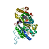
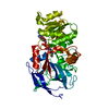
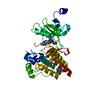
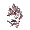
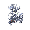
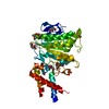
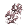
- PDB-7anj: DdahB, GDP-mannoheptose C3,5 epimerase from Campylobacter jejuni ... -
+ Open data

- Basic information
| Entry | Database: PDB / ID: 7anj | ||||||
|---|---|---|---|---|---|---|---|
| Title | DdahB, GDP-mannoheptose C3,5 epimerase from Campylobacter jejuni complexed to GDP-mannose | ||||||
 Components Components | Thymidine diphospho-4-keto-rhamnose 3,5-epimerase | ||||||
 Keywords Keywords | SUGAR BINDING PROTEIN / epimerise / sugar nucleotide / cupin fold / enzyme | ||||||
| Function / homology |  Function and homology information Function and homology informationdTDP-4-dehydrorhamnose 3,5-epimerase / dTDP-4-dehydrorhamnose 3,5-epimerase activity / polysaccharide biosynthetic process / cytosol Similarity search - Function | ||||||
| Biological species |   Campylobacter jejuni (Campylobacter) Campylobacter jejuni (Campylobacter) | ||||||
| Method |  X-RAY DIFFRACTION / MOLECULAR REPLACEMENT / Resolution: 2.35 Å X-RAY DIFFRACTION / MOLECULAR REPLACEMENT / Resolution: 2.35 Å | ||||||
 Authors Authors | Naismith, J.H. / Woodward, L. | ||||||
| Funding support |  United Kingdom, 1items United Kingdom, 1items
| ||||||
 Citation Citation | Journal: To Be Published Title: DdhaB with GDP-mannose Authors: Naismith, J.H. / Woodward, L. | ||||||
| History |
|
- Structure visualization
| Structure viewer | Molecule: MolmilJmol/JSmol |
|---|
- Downloads & links
-Download
| PDBx/mmCIF format | 7anj.cif.gz | 85.8 KB | Display | PDBx/mmCIF format |
|---|---|---|---|---|
| PDB format | pdb7anj.ent.gz | 63 KB | Display | PDB format |
| PDBx/mmJSON format | 7anj.json.gz | Tree view | PDBx/mmJSON format | |
| Others |  Other downloads Other downloads |
-Validation report
| Arichive directory | https://data.pdbj.org/pub/pdb/validation_reports/an/7anjftp://data.pdbj.org/pub/pdb/validation_reports/an/7anj | HTTPS FTP |
|---|
-Related structure data
| Related structure data |  1dzrS S: Starting model for refinement |
|---|---|
| Similar structure data |


-Links
 PDBj
PDBj- Assembly
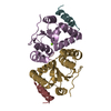
| Deposited unit | 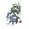
| |||||||||||||||||||||||||||
|---|---|---|---|---|---|---|---|---|---|---|---|---|---|---|---|---|---|---|---|---|---|---|---|---|---|---|---|---|
| 1 |
| |||||||||||||||||||||||||||
| Unit cell |
| |||||||||||||||||||||||||||
| Noncrystallographic symmetry (NCS) | NCS domain:
NCS domain segments:
|
-Components
| #1: Protein | Mass: 20955.885 Da / Num. of mol.: 2 Source method: isolated from a genetically manipulated source Source: (gene. exp.) Campylobacter jejuni (Campylobacter)Gene: BBR99_05345, D0W34_08620, D5I02_08520, F0H18_08555, F1P94_09915, FRS42_06315, FVZ69_07850, FW031_08545, FW918_03925, FWZ96_07945, GAX04_08395, GJ442_08190, GRS20_08795, GSH24_04195, GY415_ ...Gene: BBR99_05345, D0W34_08620, D5I02_08520, F0H18_08555, F1P94_09915, FRS42_06315, FVZ69_07850, FW031_08545, FW918_03925, FWZ96_07945, GAX04_08395, GJ442_08190, GRS20_08795, GSH24_04195, GY415_001377, GZD82_001464, HS23.15 Production host: References: UniProt: Q6EF58, dTDP-4-dehydrorhamnose 3,5-epimerase #2: Chemical | ChemComp-GDD / |   Mass: 605.341 Da / Num. of mol.: 1 / Source method: obtained synthetically / Formula: C16H25N5O16P2 Mass: 605.341 Da / Num. of mol.: 1 / Source method: obtained synthetically / Formula: C16H25N5O16P2#3: Chemical | ChemComp-GDP / |   Type: RNA linking / Mass: 443.201 Da / Num. of mol.: 1 / Source method: obtained synthetically / Formula: C10H15N5O11P2 / Comment: GDP, energy-carrying molecule*YM Type: RNA linking / Mass: 443.201 Da / Num. of mol.: 1 / Source method: obtained synthetically / Formula: C10H15N5O11P2 / Comment: GDP, energy-carrying molecule*YM#4: Water | ChemComp-HOH / |  Mass: 18.015 Da / Num. of mol.: 46 / Source method: isolated from a natural source / Formula: H2O Mass: 18.015 Da / Num. of mol.: 46 / Source method: isolated from a natural source / Formula: H2OHas ligand of interest | N | |
|---|
-Experimental details
-Experiment
| Experiment | Method: X-RAY DIFFRACTION / Number of used crystals: 1 |
|---|
- Sample preparation
| Crystal | Density Matthews: 2.06 Å3/Da / Density % sol: 40.36 % |
|---|---|
| Crystal grow | Temperature: 298 K / Method: vapor diffusion, sitting drop / Details: 24 % (w/v) PEG 1500, 20 % (v/v) glycerol |
-Data collection
| Diffraction | Mean temperature: 120 K / Serial crystal experiment: N |
|---|---|
| Diffraction source | Source: ROTATING ANODE / Type: RIGAKU MICROMAX-007 HF / Wavelength: 1.5418 Å |
| Detector | Type: RIGAKU SATURN 944 / Detector: CCD / Date: Oct 4, 2016 |
| Radiation | Protocol: SINGLE WAVELENGTH / Monochromatic (M) / Laue (L): M / Scattering type: x-ray |
| Radiation wavelength | Wavelength: 1.5418 Å / Relative weight: 1 |
| Reflection | Resolution: 2.35→53.11 Å / Num. obs: 13466 / % possible obs: 98 % / Redundancy: 3.6 % / CC1/2: 1 / Rmerge(I) obs: 0.061 / Net I/σ(I): 16.3 |
| Reflection shell | Resolution: 2.35→2.43 Å / Redundancy: 2.7 % / Rmerge(I) obs: 0.169 / Mean I/σ(I) obs: 6.1 / Num. unique obs: 879 / CC1/2: 0.9 / % possible all: 89 |
- Processing
| Software |
| ||||||||||||||||||||||||||||||||||||||||||||||||||||||||||||
|---|---|---|---|---|---|---|---|---|---|---|---|---|---|---|---|---|---|---|---|---|---|---|---|---|---|---|---|---|---|---|---|---|---|---|---|---|---|---|---|---|---|---|---|---|---|---|---|---|---|---|---|---|---|---|---|---|---|---|---|---|---|
| Refinement | Method to determine structure: MOLECULAR REPLACEMENT Starting model: 1DZR Resolution: 2.35→53.11 Å / Cor.coef. Fo:Fc: 0.931 / Cor.coef. Fo:Fc free: 0.889 / SU B: 7.741 / SU ML: 0.185 / Cross valid method: THROUGHOUT / σ(F): 0 / ESU R: 0.525 / ESU R Free: 0.269 / Stereochemistry target values: MAXIMUM LIKELIHOOD Details: HYDROGENS HAVE BEEN ADDED IN THE RIDING POSITIONS U VALUES : REFINED INDIVIDUALLY
| ||||||||||||||||||||||||||||||||||||||||||||||||||||||||||||
| Solvent computation | Ion probe radii: 0.8 Å / Shrinkage radii: 0.8 Å / VDW probe radii: 1.2 Å / Solvent model: MASK | ||||||||||||||||||||||||||||||||||||||||||||||||||||||||||||
| Displacement parameters | Biso max: 218.13 Å2 / Biso mean: 21.312 Å2 / Biso min: 2 Å2
| ||||||||||||||||||||||||||||||||||||||||||||||||||||||||||||
| Refinement step | Cycle: final / Resolution: 2.35→53.11 Å
| ||||||||||||||||||||||||||||||||||||||||||||||||||||||||||||
| Refine LS restraints |
| ||||||||||||||||||||||||||||||||||||||||||||||||||||||||||||
| Refine LS restraints NCS | Ens-ID: 1 / Number: 19512 / Refine-ID: X-RAY DIFFRACTION / Type: interatomic distance / Rms dev position: 0.08 Å / Weight position: 0.05
| ||||||||||||||||||||||||||||||||||||||||||||||||||||||||||||
| LS refinement shell | Resolution: 2.35→2.407 Å / Rfactor Rfree error: 0
|
