 Movie
Movie Controller

[English] 日本語
 Yorodumi
- PDB-7ajo: The X-ray Structure of L,D-transpeptidase LdtA from Vibrio choler... -
+ Open data

- Basic information
| Entry | Database: PDB / ID: 7ajo | ||||||
|---|---|---|---|---|---|---|---|
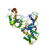
| Title | The X-ray Structure of L,D-transpeptidase LdtA from Vibrio cholerae in complex with the cross-linking reaction intermediate | ||||||
 Components Components | L,D-transpeptidase YcbB | ||||||
 Keywords Keywords | TRANSFERASE / L / D-transpeptidase | ||||||
| Function / homology |  Function and homology information Function and homology informationpeptidoglycan biosynthetic process / carboxypeptidase activity / transferase activity Similarity search - Function | ||||||
| Biological species |   Vibrio cholerae (bacteria) Vibrio cholerae (bacteria) | ||||||
| Method |  X-RAY DIFFRACTION / SYNCHROTRON / MOLECULAR REPLACEMENT / Resolution: 1.85 Å X-RAY DIFFRACTION / SYNCHROTRON / MOLECULAR REPLACEMENT / Resolution: 1.85 Å | ||||||
 Authors Authors | Batuecas, M.T. / Hermoso, J.A. | ||||||
| Funding support |  Spain, 1items Spain, 1items
| ||||||
 Citation Citation | Journal: To Be Published Title: The X-ray Structure of L,D-transpeptidase LdtA from Vibrio cholerae in complex with the cross-linking reaction intermediate Authors: Batuecas, M.T. / Hermoso, J.A. | ||||||
| History |
|
- Structure visualization
| Structure viewer | Molecule: MolmilJmol/JSmol |
|---|
- Downloads & links
-Download
| PDBx/mmCIF format | 7ajo.cif.gz | 129.1 KB | Display | PDBx/mmCIF format |
|---|---|---|---|---|
| PDB format | pdb7ajo.ent.gz | 95.6 KB | Display | PDB format |
| PDBx/mmJSON format | 7ajo.json.gz | Tree view | PDBx/mmJSON format | |
| Others |  Other downloads Other downloads |
-Validation report
| Arichive directory | https://data.pdbj.org/pub/pdb/validation_reports/aj/7ajoftp://data.pdbj.org/pub/pdb/validation_reports/aj/7ajo | HTTPS FTP |
|---|
-Related structure data
| Related structure data |  7aj9S S: Starting model for refinement |
|---|---|
| Similar structure data |


-Links
 PDBj
PDBj
- Assembly
| Deposited unit | 
| ||||||||
|---|---|---|---|---|---|---|---|---|---|
| 1 |
| ||||||||
| Unit cell |
|
-Components
| #1: Protein | Mass: 60418.012 Da / Num. of mol.: 1 Source method: isolated from a genetically manipulated source Source: (gene. exp.) Vibrio cholerae (bacteria) / Gene: ERS013138_01558, ERS013186_00077, ERS013206_00149 / Production host: |
|---|---|
| #2: Chemical | ChemComp-EDO /   Mass: 62.068 Da / Num. of mol.: 1 / Source method: obtained synthetically / Formula: C2H6O2 Mass: 62.068 Da / Num. of mol.: 1 / Source method: obtained synthetically / Formula: C2H6O2 |
| #3: Chemical | ChemComp-S2K / (  Mass: 319.311 Da / Num. of mol.: 1 / Source method: obtained synthetically / Formula: C12H21N3O7 Mass: 319.311 Da / Num. of mol.: 1 / Source method: obtained synthetically / Formula: C12H21N3O7 |
| #4: Water | ChemComp-HOH /  Mass: 18.015 Da / Num. of mol.: 466 / Source method: isolated from a natural source / Formula: H2O Mass: 18.015 Da / Num. of mol.: 466 / Source method: isolated from a natural source / Formula: H2O |
| Has ligand of interest | Y |
| Has protein modification | Y |
-Experimental details
-Experiment
| Experiment | Method: X-RAY DIFFRACTION / Number of used crystals: 1 |
|---|
- Sample preparation
| Crystal | Density Matthews: 2.93 Å3/Da / Density % sol: 58.03 % |
|---|---|
| Crystal grow | Temperature: 298 K / Method: vapor diffusion Details: 0.1 M HEPES pH 7 + 10% PEG 8000 + 10% Ethylene Glycol |
-Data collection
| Diffraction | Mean temperature: 100 K / Serial crystal experiment: N |
|---|---|
| Diffraction source | Source: SYNCHROTRON / Site: ALBA / Beamline: XALOC / Wavelength: 0.97916 Å |
| Detector | Type: DECTRIS PILATUS 6M / Detector: PIXEL / Date: Jul 19, 2017 |
| Radiation | Protocol: SINGLE WAVELENGTH / Monochromatic (M) / Laue (L): M / Scattering type: x-ray |
| Radiation wavelength | Wavelength: 0.97916 Å / Relative weight: 1 |
| Reflection | Resolution: 1.85→46.67 Å / Num. obs: 58138 / % possible obs: 99.9 % / Redundancy: 6.2 % / CC1/2: 0.997 / Rpim(I) all: 0.069 / Net I/σ(I): 9 |
| Reflection shell | Resolution: 1.85→1.89 Å / Redundancy: 6.4 % / Mean I/σ(I) obs: 6.4 / Num. unique obs: 3549 / CC1/2: 0.409 / Rpim(I) all: 1.042 / % possible all: 99.9 |
- Processing
| Software |
| ||||||||||||||||||||||||||||||||||||||||||||||||||||||||||||
|---|---|---|---|---|---|---|---|---|---|---|---|---|---|---|---|---|---|---|---|---|---|---|---|---|---|---|---|---|---|---|---|---|---|---|---|---|---|---|---|---|---|---|---|---|---|---|---|---|---|---|---|---|---|---|---|---|---|---|---|---|---|
| Refinement | Method to determine structure: MOLECULAR REPLACEMENT Starting model: 7AJ9 Resolution: 1.85→46.67 Å / Cor.coef. Fo:Fc: 0.966 / Cor.coef. Fo:Fc free: 0.95 / SU B: 3.995 / SU ML: 0.108 / Cross valid method: THROUGHOUT / σ(F): 0 / ESU R: 0.119 / ESU R Free: 0.118 / Stereochemistry target values: MAXIMUM LIKELIHOOD Details: HYDROGENS HAVE BEEN ADDED IN THE RIDING POSITIONS U VALUES : REFINED INDIVIDUALLY
| ||||||||||||||||||||||||||||||||||||||||||||||||||||||||||||
| Solvent computation | Ion probe radii: 0.8 Å / Shrinkage radii: 0.8 Å / VDW probe radii: 1.2 Å / Solvent model: MASK | ||||||||||||||||||||||||||||||||||||||||||||||||||||||||||||
| Displacement parameters | Biso max: 121.67 Å2 / Biso mean: 30.197 Å2 / Biso min: 15.11 Å2
| ||||||||||||||||||||||||||||||||||||||||||||||||||||||||||||
| Refinement step | Cycle: final / Resolution: 1.85→46.67 Å
| ||||||||||||||||||||||||||||||||||||||||||||||||||||||||||||
| Refine LS restraints |
| ||||||||||||||||||||||||||||||||||||||||||||||||||||||||||||
| LS refinement shell | Resolution: 1.85→1.898 Å / Rfactor Rfree error: 0 / Total num. of bins used: 20
|
