 Movie
Movie Controller

[English] 日本語
 Yorodumi
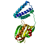
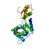
- PDB-7aie: Crystal structure of a truncated form of the KLC1-TPR domain ([A1... -
+ Open data

- Basic information
| Entry | Database: PDB / ID: 7aie | ||||||
|---|---|---|---|---|---|---|---|
| Title | Crystal structure of a truncated form of the KLC1-TPR domain ([A1-B5] fragment) - Monoclinic crystal form | ||||||
 Components Components | Kinesin light chain 1 | ||||||
 Keywords Keywords | MOTOR PROTEIN / Kinesin Light Chain / kinesin1 / TPR domain / Kif5 / accesosry protein | ||||||
| Function / homology |  Function and homology information Function and homology informationRHO GTPases activate KTN1 / Kinesins / kinesin complex / COPI-dependent Golgi-to-ER retrograde traffic / microtubule-based movement / stress granule disassembly / cytoskeletal motor activity / kinesin binding / MHC class II antigen presentation / Signaling by ALK fusions and activated point mutants ...RHO GTPases activate KTN1 / Kinesins / kinesin complex / COPI-dependent Golgi-to-ER retrograde traffic / microtubule-based movement / stress granule disassembly / cytoskeletal motor activity / kinesin binding / MHC class II antigen presentation / Signaling by ALK fusions and activated point mutants / growth cone / cytoplasmic vesicle / microtubule / cell adhesion / membrane / cytoplasm / cytosol Similarity search - Function | ||||||
| Biological species |  Homo sapiens (human) Homo sapiens (human) | ||||||
| Method |  X-RAY DIFFRACTION / SYNCHROTRON / MOLECULAR REPLACEMENT / Resolution: 3.287 Å X-RAY DIFFRACTION / SYNCHROTRON / MOLECULAR REPLACEMENT / Resolution: 3.287 Å | ||||||
 Authors Authors | Menetrey, J. / Llinas, P. | ||||||
 Citation Citation | Journal: To Be Published Title: Structural investigations of the dynamics of the TPR domain of kinesin light chain Authors: Menetrey, J. / Llinas, P. | ||||||
| History |
|
- Structure visualization
| Structure viewer | Molecule: MolmilJmol/JSmol |
|---|
- Downloads & links
-Download
| PDBx/mmCIF format | 7aie.cif.gz | 313.5 KB | Display | PDBx/mmCIF format |
|---|---|---|---|---|
| PDB format | pdb7aie.ent.gz | 256 KB | Display | PDB format |
| PDBx/mmJSON format | 7aie.json.gz | Tree view | PDBx/mmJSON format | |
| Others |  Other downloads Other downloads |
-Validation report
| Arichive directory | https://data.pdbj.org/pub/pdb/validation_reports/ai/7aieftp://data.pdbj.org/pub/pdb/validation_reports/ai/7aie | HTTPS FTP |
|---|
-Related structure data
| Related structure data |  7ai4C  5oj8S S: Starting model for refinement C: citing same article ( |
|---|---|
| Similar structure data |


-Links
 PDBj
PDBj


- Assembly
| Deposited unit | 
| ||||||||
|---|---|---|---|---|---|---|---|---|---|
| 1 | 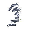
| ||||||||
| 2 | 
| ||||||||
| 3 | 
| ||||||||
| 4 | 
| ||||||||
| Unit cell |
|
-Components
| #1: Protein | Mass: 28577.311 Da / Num. of mol.: 4 Source method: isolated from a genetically manipulated source Source: (gene. exp.) Homo sapiens (human) / Gene: KLC1, KLC, KNS2 / Production host:  #2: Chemical |   Mass: 24.305 Da / Num. of mol.: 2 / Source method: obtained synthetically / Formula: Mg Mass: 24.305 Da / Num. of mol.: 2 / Source method: obtained synthetically / Formula: MgHas ligand of interest | N | |
|---|
-Experimental details
-Experiment
| Experiment | Method: X-RAY DIFFRACTION / Number of used crystals: 1 |
|---|
- Sample preparation
| Crystal | Density Matthews: 2.52 Å3/Da / Density % sol: 51.21 % |
|---|---|
| Crystal grow | Temperature: 290 K / Method: vapor diffusion, hanging drop Details: 20% PEG8000, 0.6M magnesium acetate, 50mM sodium cacodylate pH 6.5 |
-Data collection
| Diffraction | Mean temperature: 100 K / Serial crystal experiment: N |
|---|---|
| Diffraction source | Source: SYNCHROTRON / Site: SOLEIL  / Beamline: PROXIMA 1 / Wavelength: 0.97857 Å / Beamline: PROXIMA 1 / Wavelength: 0.97857 Å |
| Detector | Type: DECTRIS EIGER X 16M / Detector: PIXEL / Date: Sep 14, 2017 |
| Radiation | Protocol: SINGLE WAVELENGTH / Monochromatic (M) / Laue (L): M / Scattering type: x-ray |
| Radiation wavelength | Wavelength: 0.97857 Å / Relative weight: 1 |
| Reflection | Resolution: 3.287→46.33 Å / Num. obs: 17338 / % possible obs: 98.6 % / Redundancy: 3.7 % / CC1/2: 0.991 / Net I/σ(I): 4.55 |
| Reflection shell | Resolution: 3.287→3.48 Å / Mean I/σ(I) obs: 0.93 / Num. unique obs: 2626 / CC1/2: 0.537 |
- Processing
| Software |
| |||||||||||||||||||||||||||||||||||||||||||||||||||||||||||||||||||||||||||||||||||||||||||||||||||||||||||||||||||||||||||||
|---|---|---|---|---|---|---|---|---|---|---|---|---|---|---|---|---|---|---|---|---|---|---|---|---|---|---|---|---|---|---|---|---|---|---|---|---|---|---|---|---|---|---|---|---|---|---|---|---|---|---|---|---|---|---|---|---|---|---|---|---|---|---|---|---|---|---|---|---|---|---|---|---|---|---|---|---|---|---|---|---|---|---|---|---|---|---|---|---|---|---|---|---|---|---|---|---|---|---|---|---|---|---|---|---|---|---|---|---|---|---|---|---|---|---|---|---|---|---|---|---|---|---|---|---|---|---|
| Refinement | Method to determine structure: MOLECULAR REPLACEMENT Starting model: 5OJ8 Resolution: 3.287→46.33 Å / Cor.coef. Fo:Fc: 0.941 / Cor.coef. Fo:Fc free: 0.878 / Cross valid method: THROUGHOUT / σ(F): 0 / SU Rfree Blow DPI: 0.467
| |||||||||||||||||||||||||||||||||||||||||||||||||||||||||||||||||||||||||||||||||||||||||||||||||||||||||||||||||||||||||||||
| Displacement parameters | Biso max: 168.66 Å2 / Biso mean: 116.08 Å2 / Biso min: 62.05 Å2
| |||||||||||||||||||||||||||||||||||||||||||||||||||||||||||||||||||||||||||||||||||||||||||||||||||||||||||||||||||||||||||||
| Refine analyze | Luzzati coordinate error obs: 0.53 Å | |||||||||||||||||||||||||||||||||||||||||||||||||||||||||||||||||||||||||||||||||||||||||||||||||||||||||||||||||||||||||||||
| Refinement step | Cycle: final / Resolution: 3.287→46.33 Å
| |||||||||||||||||||||||||||||||||||||||||||||||||||||||||||||||||||||||||||||||||||||||||||||||||||||||||||||||||||||||||||||
| Refine LS restraints |
| |||||||||||||||||||||||||||||||||||||||||||||||||||||||||||||||||||||||||||||||||||||||||||||||||||||||||||||||||||||||||||||
| LS refinement shell | Resolution: 3.29→3.32 Å / Rfactor Rfree error: 0 / Total num. of bins used: 44
| |||||||||||||||||||||||||||||||||||||||||||||||||||||||||||||||||||||||||||||||||||||||||||||||||||||||||||||||||||||||||||||
| Refinement TLS params. | Method: refined / Refine-ID: X-RAY DIFFRACTION
| |||||||||||||||||||||||||||||||||||||||||||||||||||||||||||||||||||||||||||||||||||||||||||||||||||||||||||||||||||||||||||||
| Refinement TLS group |
|
