 Movie
Movie Controller

[English] 日本語
 Yorodumi
- PDB-7agh: Crystal structure of SF kinase YihV from E. coli in complex with ... -
+ Open data

- Basic information
| Entry | Database: PDB / ID: 7agh | ||||||
|---|---|---|---|---|---|---|---|
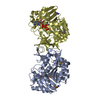
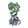
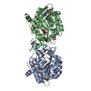
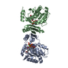
| Title | Crystal structure of SF kinase YihV from E. coli in complex with AMPPNP-Mg | ||||||
 Components Components | Sulfofructose kinase | ||||||
 Keywords Keywords | TRANSFERASE / sulfofructose / SF kinase / carbohydrate kinase / sulfoglycolysis | ||||||
| Function / homology |  Function and homology information Function and homology informationsulfofructose kinase / 6-deoxy-6-sulfofructose kinase activity / : / 6-sulfoquinovose(1-) catabolic process / carbohydrate phosphorylation / kinase activity / ATP binding / cytosol Similarity search - Function | ||||||
| Biological species |  | ||||||
| Method |  X-RAY DIFFRACTION / SYNCHROTRON / MOLECULAR REPLACEMENT / Resolution: 2.93 Å X-RAY DIFFRACTION / SYNCHROTRON / MOLECULAR REPLACEMENT / Resolution: 2.93 Å | ||||||
 Authors Authors | Sharma, M. / Davies, G.J. / Jin, Y. | ||||||
| Funding support |  United Kingdom, 1items United Kingdom, 1items
| ||||||
 Citation Citation | Journal: Acs Cent.Sci. / Year: 2021 Title: Molecular Basis of Sulfosugar Selectivity in Sulfoglycolysis. Authors: Sharma, M. / Abayakoon, P. / Epa, R. / Jin, Y. / Lingford, J.P. / Shimada, T. / Nakano, M. / Mui, J.W. / Ishihama, A. / Goddard-Borger, E.D. / Davies, G.J. / Williams, S.J. | ||||||
| History |
|
- Structure visualization
| Structure viewer | Molecule: MolmilJmol/JSmol |
|---|
- Downloads & links
-Download
| PDBx/mmCIF format | 7agh.cif.gz | 224.9 KB | Display | PDBx/mmCIF format |
|---|---|---|---|---|
| PDB format | pdb7agh.ent.gz | 176.8 KB | Display | PDB format |
| PDBx/mmJSON format | 7agh.json.gz | Tree view | PDBx/mmJSON format | |
| Others |  Other downloads Other downloads |
-Validation report
| Arichive directory | https://data.pdbj.org/pub/pdb/validation_reports/ag/7aghftp://data.pdbj.org/pub/pdb/validation_reports/ag/7agh | HTTPS FTP |
|---|
-Related structure data
| Related structure data | 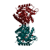 7ag1C 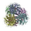 7ag4C 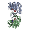 7ag6SC  7ag7C 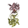 7agkC 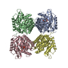 7ne2C S: Starting model for refinement C: citing same article ( |
|---|---|
| Similar structure data |


-Links
 PDBj
PDBj


- Assembly
| Deposited unit | 
| ||||||||||||||||||||||||||||||||||||||||||||||||||||||||||||||||||||||||||||||||||||||||||||||||||
|---|---|---|---|---|---|---|---|---|---|---|---|---|---|---|---|---|---|---|---|---|---|---|---|---|---|---|---|---|---|---|---|---|---|---|---|---|---|---|---|---|---|---|---|---|---|---|---|---|---|---|---|---|---|---|---|---|---|---|---|---|---|---|---|---|---|---|---|---|---|---|---|---|---|---|---|---|---|---|---|---|---|---|---|---|---|---|---|---|---|---|---|---|---|---|---|---|---|---|---|
| 1 | 
| ||||||||||||||||||||||||||||||||||||||||||||||||||||||||||||||||||||||||||||||||||||||||||||||||||
| 2 | 
| ||||||||||||||||||||||||||||||||||||||||||||||||||||||||||||||||||||||||||||||||||||||||||||||||||
| Unit cell |
| ||||||||||||||||||||||||||||||||||||||||||||||||||||||||||||||||||||||||||||||||||||||||||||||||||
| Noncrystallographic symmetry (NCS) | NCS domain:
NCS domain segments: Component-ID: _ / Beg auth comp-ID: MET / Beg label comp-ID: MET / End auth comp-ID: LEU / End label comp-ID: LEU / Refine code: _ / Auth seq-ID: 1 - 296 / Label seq-ID: 1 - 296
NCS ensembles :
|
-Components
| #1: Protein | Mass: 32829.723 Da / Num. of mol.: 4 Source method: isolated from a genetically manipulated source Source: (gene. exp.) Strain: K12 / Gene: yihV, b3883, JW5568 / Plasmid: pet21b / Production host: #2: Chemical | ChemComp-ANP /   Mass: 506.196 Da / Num. of mol.: 4 / Source method: obtained synthetically / Formula: C10H17N6O12P3 / Feature type: SUBJECT OF INVESTIGATION / Comment: AMP-PNP, energy-carrying molecule analogue*YM Mass: 506.196 Da / Num. of mol.: 4 / Source method: obtained synthetically / Formula: C10H17N6O12P3 / Feature type: SUBJECT OF INVESTIGATION / Comment: AMP-PNP, energy-carrying molecule analogue*YM#3: Chemical |   Mass: 24.305 Da / Num. of mol.: 3 / Source method: obtained synthetically / Formula: Mg / Feature type: SUBJECT OF INVESTIGATION Mass: 24.305 Da / Num. of mol.: 3 / Source method: obtained synthetically / Formula: Mg / Feature type: SUBJECT OF INVESTIGATION#4: Water | ChemComp-HOH / |  Mass: 18.015 Da / Num. of mol.: 36 / Source method: isolated from a natural source / Formula: H2O Mass: 18.015 Da / Num. of mol.: 36 / Source method: isolated from a natural source / Formula: H2OHas ligand of interest | Y | |
|---|
-Experimental details
-Experiment
| Experiment | Method: X-RAY DIFFRACTION / Number of used crystals: 1 |
|---|
- Sample preparation
| Crystal | Density Matthews: 2.2 Å3/Da / Density % sol: 44.01 % |
|---|---|
| Crystal grow | Temperature: 293 K / Method: vapor diffusion, sitting drop / pH: 5.5 Details: 25% PEG 3350w/v, 0.2 M MgCl2.6H2O, 0.1 M BisTris pH5.5 |
-Data collection
| Diffraction | Mean temperature: 100 K / Serial crystal experiment: N | ||||||||||||||||||||||||||||||
|---|---|---|---|---|---|---|---|---|---|---|---|---|---|---|---|---|---|---|---|---|---|---|---|---|---|---|---|---|---|---|---|
| Diffraction source | Source: SYNCHROTRON / Site: Diamond / Beamline: I04 / Wavelength: 0.9795 Å | ||||||||||||||||||||||||||||||
| Detector | Type: DECTRIS EIGER X 16M / Detector: PIXEL / Date: Feb 12, 2017 | ||||||||||||||||||||||||||||||
| Radiation | Protocol: SINGLE WAVELENGTH / Monochromatic (M) / Laue (L): M / Scattering type: x-ray | ||||||||||||||||||||||||||||||
| Radiation wavelength | Wavelength: 0.9795 Å / Relative weight: 1 | ||||||||||||||||||||||||||||||
| Reflection | Resolution: 2.93→59.21 Å / Num. obs: 25634 / % possible obs: 100 % / Redundancy: 6.5 % / CC1/2: 0.995 / Rmerge(I) obs: 0.119 / Rpim(I) all: 0.05 / Rrim(I) all: 0.129 / Net I/σ(I): 10.4 / Num. measured all: 165747 / Scaling rejects: 335 | ||||||||||||||||||||||||||||||
| Reflection shell | Diffraction-ID: 1
|
- Processing
| Software |
| |||||||||||||||||||||||||||||||||||||||||||||||||||||||||||||||||
|---|---|---|---|---|---|---|---|---|---|---|---|---|---|---|---|---|---|---|---|---|---|---|---|---|---|---|---|---|---|---|---|---|---|---|---|---|---|---|---|---|---|---|---|---|---|---|---|---|---|---|---|---|---|---|---|---|---|---|---|---|---|---|---|---|---|---|
| Refinement | Method to determine structure: MOLECULAR REPLACEMENT Starting model: 7AG6 Resolution: 2.93→59.01 Å / Cor.coef. Fo:Fc: 0.923 / Cor.coef. Fo:Fc free: 0.89 / SU B: 17.156 / SU ML: 0.324 / Cross valid method: THROUGHOUT / σ(F): 0 / ESU R Free: 0.453 / Stereochemistry target values: MAXIMUM LIKELIHOOD Details: HYDROGENS HAVE BEEN ADDED IN THE RIDING POSITIONS U VALUES : REFINED INDIVIDUALLY
| |||||||||||||||||||||||||||||||||||||||||||||||||||||||||||||||||
| Solvent computation | Ion probe radii: 0.8 Å / Shrinkage radii: 0.8 Å / VDW probe radii: 1.2 Å / Solvent model: MASK | |||||||||||||||||||||||||||||||||||||||||||||||||||||||||||||||||
| Displacement parameters | Biso max: 102.55 Å2 / Biso mean: 39.025 Å2 / Biso min: 0.68 Å2
| |||||||||||||||||||||||||||||||||||||||||||||||||||||||||||||||||
| Refinement step | Cycle: final / Resolution: 2.93→59.01 Å
| |||||||||||||||||||||||||||||||||||||||||||||||||||||||||||||||||
| Refine LS restraints |
| |||||||||||||||||||||||||||||||||||||||||||||||||||||||||||||||||
| Refine LS restraints NCS | Refine-ID: X-RAY DIFFRACTION / Type: interatomic distance / Weight position: 0.05
| |||||||||||||||||||||||||||||||||||||||||||||||||||||||||||||||||
| LS refinement shell | Resolution: 2.93→3.006 Å / Rfactor Rfree error: 0 / Total num. of bins used: 20
|
