 Movie
Movie Controller


+ Open data

- Basic information
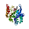
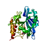
| Entry | Database: PDB / ID: 7aez | |||||||||
|---|---|---|---|---|---|---|---|---|---|---|
| Title | Crystal structure of the metallo-beta-lactamase NDM-7 with 407 | |||||||||
 Components Components | Metallo-beta-lactamase NDM-7 | |||||||||
 Keywords Keywords | HYDROLASE / HYDROLASE METALLO-BETA-LACTAMASE ANTIBIOTIC RESISTANCE | |||||||||
| Function / homology |  Function and homology information Function and homology informationantibiotic catabolic process / beta-lactamase / hydrolase activity / metal ion binding Similarity search - Function | |||||||||
| Biological species |  Klebsiella pneumoniae (bacteria) Klebsiella pneumoniae (bacteria) | |||||||||
| Method |  X-RAY DIFFRACTION / SYNCHROTRON / MOLECULAR REPLACEMENT / molecular replacement / Resolution: 1.018 Å X-RAY DIFFRACTION / SYNCHROTRON / MOLECULAR REPLACEMENT / molecular replacement / Resolution: 1.018 Å | |||||||||
 Authors Authors | Brem, J. / Schofield, C.J. | |||||||||
| Funding support |  United Kingdom, 2items United Kingdom, 2items
| |||||||||
 Citation Citation | Journal: To Be Published Title: Not available yet Authors: Brem, J. / Schofield, C.J. | |||||||||
| History |
|
- Structure visualization
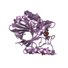
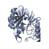
| Structure viewer | Molecule: MolmilJmol/JSmol |
|---|
- Downloads & links
-Download
| PDBx/mmCIF format | 7aez.cif.gz | 165.1 KB | Display | PDBx/mmCIF format |
|---|---|---|---|---|
| PDB format | pdb7aez.ent.gz | 132 KB | Display | PDB format |
| PDBx/mmJSON format | 7aez.json.gz | Tree view | PDBx/mmJSON format | |
| Others |  Other downloads Other downloads |
-Validation report
| Arichive directory | https://data.pdbj.org/pub/pdb/validation_reports/ae/7aezftp://data.pdbj.org/pub/pdb/validation_reports/ae/7aez | HTTPS FTP |
|---|
-Related structure data
| Related structure data | 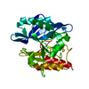 4tzfS S: Starting model for refinement |
|---|---|
| Similar structure data |

-Links
 PDBj
PDBj

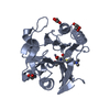
- Assembly
| Deposited unit | 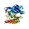
| ||||||||
|---|---|---|---|---|---|---|---|---|---|
| 1 |
| ||||||||
| Unit cell |
|
-Components
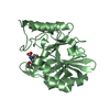
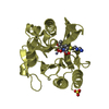
-Protein , 1 types, 1 molecules A
| #1: Protein | Mass: 24400.400 Da / Num. of mol.: 1 Source method: isolated from a genetically manipulated source Details: CYS208 is sp2 hybridised at CA / Source: (gene. exp.) Klebsiella pneumoniae (bacteria) / Gene: blaNDM-7 / Production host: |
|---|
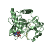
-Non-polymers , 7 types, 340 molecules 


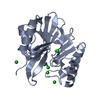
| #2: Chemical | ChemComp-R8W /  Mass: 360.409 Da / Num. of mol.: 1 / Source method: obtained synthetically / Formula: C21H20N4O2 / Feature type: SUBJECT OF INVESTIGATION Mass: 360.409 Da / Num. of mol.: 1 / Source method: obtained synthetically / Formula: C21H20N4O2 / Feature type: SUBJECT OF INVESTIGATION | ||||||||||
|---|---|---|---|---|---|---|---|---|---|---|---|
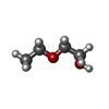
| #3: Chemical |  Mass: 65.409 Da / Num. of mol.: 2 / Source method: obtained synthetically / Formula: Zn Mass: 65.409 Da / Num. of mol.: 2 / Source method: obtained synthetically / Formula: Zn#4: Chemical |  Mass: 92.094 Da / Num. of mol.: 2 / Source method: obtained synthetically / Formula: C3H8O3 Mass: 92.094 Da / Num. of mol.: 2 / Source method: obtained synthetically / Formula: C3H8O3#5: Chemical | ChemComp-EDO / |  Mass: 62.068 Da / Num. of mol.: 1 / Source method: obtained synthetically / Formula: C2H6O2 Mass: 62.068 Da / Num. of mol.: 1 / Source method: obtained synthetically / Formula: C2H6O2#6: Chemical | ChemComp-NA / |  Mass: 22.990 Da / Num. of mol.: 1 / Source method: obtained synthetically / Formula: Na Mass: 22.990 Da / Num. of mol.: 1 / Source method: obtained synthetically / Formula: Na#7: Chemical | ChemComp-ETX / |  Mass: 90.121 Da / Num. of mol.: 1 / Source method: obtained synthetically / Formula: C4H10O2 Mass: 90.121 Da / Num. of mol.: 1 / Source method: obtained synthetically / Formula: C4H10O2#8: Water | ChemComp-HOH / | Mass: 18.015 Da / Num. of mol.: 332 / Source method: isolated from a natural source / Formula: H2O |
-Details
| Has ligand of interest | Y |
|---|
-Experimental details
-Experiment
| Experiment | Method: X-RAY DIFFRACTION / Number of used crystals: 1 |
|---|
- Sample preparation
| Crystal | Density Matthews: 2.11 Å3/Da / Density % sol: 41.82 % |
|---|---|
| Crystal grow | Temperature: 293 K / Method: vapor diffusion, sitting drop / pH: 5.5 / Details: 0.1M Bis Tris pH 5.5 25% PEG3350 |
-Data collection
| Diffraction | Mean temperature: 100 K / Serial crystal experiment: N | ||||||||||||||||||||||||||||||
|---|---|---|---|---|---|---|---|---|---|---|---|---|---|---|---|---|---|---|---|---|---|---|---|---|---|---|---|---|---|---|---|
| Diffraction source | Source: SYNCHROTRON / Site: Diamond / Beamline: I03 / Wavelength: 0.9763 Å | ||||||||||||||||||||||||||||||
| Detector | Type: DECTRIS PILATUS 6M / Detector: PIXEL / Date: May 7, 2017 | ||||||||||||||||||||||||||||||
| Radiation | Protocol: SINGLE WAVELENGTH / Monochromatic (M) / Laue (L): M / Scattering type: x-ray | ||||||||||||||||||||||||||||||
| Radiation wavelength | Wavelength: 0.9763 Å / Relative weight: 1 | ||||||||||||||||||||||||||||||
| Reflection | Resolution: 1.01843→27.97 Å / Num. obs: 97817 / % possible obs: 92.2 % / Redundancy: 11.4 % / Biso Wilson estimate: 8.56 Å2 / CC1/2: 1 / Rmerge(I) obs: 0.057 / Rpim(I) all: 0.017 / Rrim(I) all: 0.06 / Net I/σ(I): 24.8 | ||||||||||||||||||||||||||||||
| Reflection shell | Diffraction-ID: 1
|
-Phasing
| Phasing | Method: molecular replacement | |||||||||
|---|---|---|---|---|---|---|---|---|---|---|
| Phasing MR |
|
- Processing
| Software |
| ||||||||||||||||||||||||||||||||||||||||||||||||||||||||||||||||||||||||||||||||||||||||||
|---|---|---|---|---|---|---|---|---|---|---|---|---|---|---|---|---|---|---|---|---|---|---|---|---|---|---|---|---|---|---|---|---|---|---|---|---|---|---|---|---|---|---|---|---|---|---|---|---|---|---|---|---|---|---|---|---|---|---|---|---|---|---|---|---|---|---|---|---|---|---|---|---|---|---|---|---|---|---|---|---|---|---|---|---|---|---|---|---|---|---|---|
| Refinement | Method to determine structure: MOLECULAR REPLACEMENT Starting model: 4TZF Resolution: 1.018→27.719 Å / SU ML: 0.06 / Cross valid method: THROUGHOUT / σ(F): 1.36 / Phase error: 9.59 / Stereochemistry target values: ML
| ||||||||||||||||||||||||||||||||||||||||||||||||||||||||||||||||||||||||||||||||||||||||||
| Solvent computation | Shrinkage radii: 0.9 Å / VDW probe radii: 1.11 Å / Solvent model: FLAT BULK SOLVENT MODEL | ||||||||||||||||||||||||||||||||||||||||||||||||||||||||||||||||||||||||||||||||||||||||||
| Displacement parameters | Biso max: 50.2 Å2 / Biso mean: 13.1449 Å2 / Biso min: 6.05 Å2 | ||||||||||||||||||||||||||||||||||||||||||||||||||||||||||||||||||||||||||||||||||||||||||
| Refinement step | Cycle: final / Resolution: 1.018→27.719 Å
| ||||||||||||||||||||||||||||||||||||||||||||||||||||||||||||||||||||||||||||||||||||||||||
| Refine LS restraints |
| ||||||||||||||||||||||||||||||||||||||||||||||||||||||||||||||||||||||||||||||||||||||||||
| LS refinement shell | Refine-ID: X-RAY DIFFRACTION / Rfactor Rfree error: 0
|
