 Movie
Movie Controller


+ Open data

- Basic information
| Entry | Database: PDB / ID: 7aal | ||||||
|---|---|---|---|---|---|---|---|
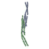
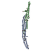
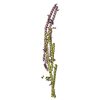
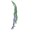
| Title | Crystal structure of the F-BAR domain of PSTIPIP1, G258A mutant | ||||||
 Components Components | Proline-serine-threonine phosphatase-interacting protein 1 | ||||||
 Keywords Keywords | SIGNALING PROTEIN / pyogenic arthritis / pyoderma gangrenosum and acne (PAPA) / Inflammatory response / membrane binding | ||||||
| Function / homology |  Function and homology information Function and homology informationuropod / cleavage furrow / The NLRP3 inflammasome / Purinergic signaling in leishmaniasis infection / endocytosis / lamellipodium / cytoskeleton / cell adhesion / inflammatory response / innate immune response ...uropod / cleavage furrow / The NLRP3 inflammasome / Purinergic signaling in leishmaniasis infection / endocytosis / lamellipodium / cytoskeleton / cell adhesion / inflammatory response / innate immune response / perinuclear region of cytoplasm / signal transduction / membrane / identical protein binding / plasma membrane / cytoplasm / cytosol Similarity search - Function | ||||||
| Biological species |  Homo sapiens (human) Homo sapiens (human) | ||||||
| Method |  X-RAY DIFFRACTION / SYNCHROTRON / MOLECULAR REPLACEMENT / Resolution: 1.97 Å X-RAY DIFFRACTION / SYNCHROTRON / MOLECULAR REPLACEMENT / Resolution: 1.97 Å | ||||||
 Authors Authors | Manso, J.A. / Alcon, P. / Bayon, Y. / Alonso, A. / de Pereda, J.M. | ||||||
| Funding support |  Spain, 1items Spain, 1items
| ||||||
 Citation Citation | Journal: Cell.Mol.Life Sci. / Year: 2022 Title: PSTPIP1-LYP phosphatase interaction: structural basis and implications for autoinflammatory disorders. Authors: Manso, J.A. / Marcos, T. / Ruiz-Martin, V. / Casas, J. / Alcon, P. / Sanchez Crespo, M. / Bayon, Y. / de Pereda, J.M. / Alonso, A. | ||||||
| History |
|
- Structure visualization
| Structure viewer | Molecule: MolmilJmol/JSmol |
|---|
- Downloads & links
-Download
| PDBx/mmCIF format | 7aal.cif.gz | 293.7 KB | Display | PDBx/mmCIF format |
|---|---|---|---|---|
| PDB format | pdb7aal.ent.gz | 197.5 KB | Display | PDB format |
| PDBx/mmJSON format | 7aal.json.gz | Tree view | PDBx/mmJSON format | |
| Others |  Other downloads Other downloads |
-Validation report
| Arichive directory | https://data.pdbj.org/pub/pdb/validation_reports/aa/7aalftp://data.pdbj.org/pub/pdb/validation_reports/aa/7aal | HTTPS FTP |
|---|
-Related structure data
| Related structure data |  7aamC  7aanC  4wpeS C: citing same article ( S: Starting model for refinement |
|---|---|
| Similar structure data |

-Links
 PDBj
PDBj


- Assembly
| Deposited unit | 
| |||||||||||||||||||||||||||||||||||||||||||||||||||||||||||||||||||||||||||||||||||||||
|---|---|---|---|---|---|---|---|---|---|---|---|---|---|---|---|---|---|---|---|---|---|---|---|---|---|---|---|---|---|---|---|---|---|---|---|---|---|---|---|---|---|---|---|---|---|---|---|---|---|---|---|---|---|---|---|---|---|---|---|---|---|---|---|---|---|---|---|---|---|---|---|---|---|---|---|---|---|---|---|---|---|---|---|---|---|---|---|---|
| 1 |
| |||||||||||||||||||||||||||||||||||||||||||||||||||||||||||||||||||||||||||||||||||||||
| Unit cell |
| |||||||||||||||||||||||||||||||||||||||||||||||||||||||||||||||||||||||||||||||||||||||
| Noncrystallographic symmetry (NCS) | NCS domain:
NCS domain segments: Ens-ID: 1
|