 Movie
Movie Controller

[English] 日本語
 Yorodumi
- PDB-7aa2: Chaetomium thermophilum FAD-dependent oxidoreductase in complex w... -
+ Open data

- Basic information
| Entry | Database: PDB / ID: 7aa2 | |||||||||||||||||||||
|---|---|---|---|---|---|---|---|---|---|---|---|---|---|---|---|---|---|---|---|---|---|---|
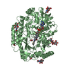
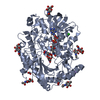
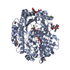
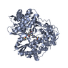
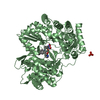
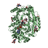
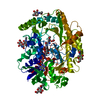
| Title | Chaetomium thermophilum FAD-dependent oxidoreductase in complex with ABTS | |||||||||||||||||||||
 Components Components | FAD-dependent oxidoreductase | |||||||||||||||||||||
 Keywords Keywords | OXIDOREDUCTASE / Chaetomium thermophilum / glucose-methanol-choline oxidoreductase / ABTS / co-crystallization | |||||||||||||||||||||
| Function / homology | Chem-EBS / DIHYDROFLAVINE-ADENINE DINUCLEOTIDE / FORMIC ACID Function and homology information Function and homology information | |||||||||||||||||||||
| Biological species |  Chaetomium thermophilum var. thermophilum DSM 1495 (fungus) Chaetomium thermophilum var. thermophilum DSM 1495 (fungus) | |||||||||||||||||||||
| Method |  X-RAY DIFFRACTION / SYNCHROTRON / MOLECULAR REPLACEMENT / Resolution: 1.4 Å X-RAY DIFFRACTION / SYNCHROTRON / MOLECULAR REPLACEMENT / Resolution: 1.4 Å | |||||||||||||||||||||
 Authors Authors | Svecova, L. / Skalova, T. / Kolenko, P. / Koval, T. / Oestergaard, L.H. / Dohnalek, J. | |||||||||||||||||||||
| Funding support |  Czech Republic, 6items Czech Republic, 6items
| |||||||||||||||||||||
 Citation Citation | Journal: Acta Crystallogr D Struct Biol / Year: 2021 Title: Crystallographic fragment screening-based study of a novel FAD-dependent oxidoreductase from Chaetomium thermophilum. Authors: Svecova, L. / Ostergaard, L.H. / Skalova, T. / Schnorr, K.M. / Koval', T. / Kolenko, P. / Stransky, J. / Sedlak, D. / Duskova, J. / Trundova, M. / Hasek, J. / Dohnalek, J. | |||||||||||||||||||||
| History |
|
- Structure visualization
| Structure viewer | Molecule: MolmilJmol/JSmol |
|---|
- Downloads & links
-Download
| PDBx/mmCIF format | 7aa2.cif.gz | 493.9 KB | Display | PDBx/mmCIF format |
|---|---|---|---|---|
| PDB format | pdb7aa2.ent.gz | 386.6 KB | Display | PDB format |
| PDBx/mmJSON format | 7aa2.json.gz | Tree view | PDBx/mmJSON format | |
| Others |  Other downloads Other downloads |
-Validation report
| Arichive directory | https://data.pdbj.org/pub/pdb/validation_reports/aa/7aa2ftp://data.pdbj.org/pub/pdb/validation_reports/aa/7aa2 | HTTPS FTP |
|---|
-Related structure data
| Related structure data |  6ze2SC  6ze3C  6ze4C  6ze5C  6ze6C  6ze7C S: Starting model for refinement C: citing same article ( |
|---|---|
| Similar structure data | |
| Experimental dataset #1 | Data reference: 10.15785/SBGRID/810 / Data set type: diffraction image data |
-Links
 PDBj
PDBj- Assembly
| Deposited unit | 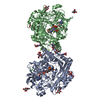
| ||||||||
|---|---|---|---|---|---|---|---|---|---|
| 1 | 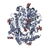
| ||||||||
| 2 | 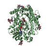
| ||||||||
| Unit cell |
|
-Components
-Protein , 1 types, 2 molecules AB
| #1: Protein | Mass: 64267.230 Da / Num. of mol.: 2 Source method: isolated from a genetically manipulated source Details: The provided sequence corresponds to the mass spectrometry analysis of sample used for crystallization Source: (gene. exp.) Chaetomium thermophilum var. thermophilum DSM 1495 (fungus)Strain: DSM 1495 / Gene: CTHT_0048040 / Production host: |
|---|
-Sugars , 2 types, 11 molecules 
| #2: Polysaccharide | Source method: isolated from a genetically manipulated source #4: Sugar | ChemComp-NAG /  Type: D-saccharide, beta linking / Mass: 221.208 Da / Num. of mol.: 9 / Source method: obtained synthetically / Formula: C8H15NO6 Type: D-saccharide, beta linking / Mass: 221.208 Da / Num. of mol.: 9 / Source method: obtained synthetically / Formula: C8H15NO6 |
|---|
-Non-polymers , 6 types, 1663 molecules 


| #3: Chemical |  Mass: 787.566 Da / Num. of mol.: 2 / Source method: obtained synthetically / Formula: C27H35N9O15P2 / Feature type: SUBJECT OF INVESTIGATION Mass: 787.566 Da / Num. of mol.: 2 / Source method: obtained synthetically / Formula: C27H35N9O15P2 / Feature type: SUBJECT OF INVESTIGATION#5: Chemical |  Mass: 514.619 Da / Num. of mol.: 2 / Source method: obtained synthetically / Formula: C18H18N4O6S4 / Feature type: SUBJECT OF INVESTIGATION Mass: 514.619 Da / Num. of mol.: 2 / Source method: obtained synthetically / Formula: C18H18N4O6S4 / Feature type: SUBJECT OF INVESTIGATION#6: Chemical |  Mass: 46.025 Da / Num. of mol.: 2 / Source method: obtained synthetically / Formula: CH2O2 Mass: 46.025 Da / Num. of mol.: 2 / Source method: obtained synthetically / Formula: CH2O2#7: Chemical |  Mass: 24.305 Da / Num. of mol.: 3 / Source method: obtained synthetically / Formula: Mg Mass: 24.305 Da / Num. of mol.: 3 / Source method: obtained synthetically / Formula: Mg#8: Chemical | ChemComp-CL / |  Mass: 35.453 Da / Num. of mol.: 1 / Source method: obtained synthetically / Formula: Cl Mass: 35.453 Da / Num. of mol.: 1 / Source method: obtained synthetically / Formula: Cl#9: Water | ChemComp-HOH / | Mass: 18.015 Da / Num. of mol.: 1653 / Source method: isolated from a natural source / Formula: H2O |
|---|
-Details
| Has ligand of interest | Y |
|---|---|
| Has protein modification | Y |
-Experimental details
-Experiment
| Experiment | Method: X-RAY DIFFRACTION / Number of used crystals: 1 |
|---|
- Sample preparation
| Crystal | Density Matthews: 2.36 Å3/Da / Density % sol: 47.9 % |
|---|---|
| Crystal grow | Temperature: 293.15 K / Method: vapor diffusion, hanging drop / pH: 5.5 Details: 17 % (w/v) PEG MME 5000, 0.1 M sodium acetate, pH 5.5, 0.16 M magnesium formate, 20 mM MgCl2, 8.5 mM 2,2'-azino-bis(3-ethylbenzothiazoline-6-sulfonic acid), protein concentration 8 mg/ml |
-Data collection
| Diffraction | Mean temperature: 100 K / Serial crystal experiment: N |
|---|---|
| Diffraction source | Source: SYNCHROTRON / Site: PETRA III, EMBL c/o DESY  / Beamline: P13 (MX1) / Wavelength: 0.97626 Å / Beamline: P13 (MX1) / Wavelength: 0.97626 Å |
| Detector | Type: DECTRIS PILATUS 6M / Detector: PIXEL / Date: Jun 24, 2019 |
| Radiation | Protocol: SINGLE WAVELENGTH / Monochromatic (M) / Laue (L): M / Scattering type: x-ray |
| Radiation wavelength | Wavelength: 0.97626 Å / Relative weight: 1 |
| Reflection | Resolution: 1.4→49.343 Å / Num. obs: 223121 / % possible obs: 95.5 % / Observed criterion σ(I): -3.7 / Redundancy: 3.7 % / Biso Wilson estimate: 16.2 Å2 / CC1/2: 0.999 / Rmerge(I) obs: 0.058 / Rpim(I) all: 0.033 / Rrim(I) all: 0.067 / Net I/σ(I): 9.2 |
| Reflection shell | Resolution: 1.4→1.42 Å / Redundancy: 3.8 % / Rmerge(I) obs: 0.925 / Mean I/σ(I) obs: 1.4 / Num. unique obs: 11468 / CC1/2: 0.589 / Rpim(I) all: 0.542 / Rrim(I) all: 1.079 / % possible all: 99.8 |
- Processing
| Software |
| ||||||||||||||||||||||||||||||||||||||||||||||||||||||||||||||||||||||||||||||||||||||||||||||||||||||||||||||||||||||||||||||||||||||||||||||||||||||
|---|---|---|---|---|---|---|---|---|---|---|---|---|---|---|---|---|---|---|---|---|---|---|---|---|---|---|---|---|---|---|---|---|---|---|---|---|---|---|---|---|---|---|---|---|---|---|---|---|---|---|---|---|---|---|---|---|---|---|---|---|---|---|---|---|---|---|---|---|---|---|---|---|---|---|---|---|---|---|---|---|---|---|---|---|---|---|---|---|---|---|---|---|---|---|---|---|---|---|---|---|---|---|---|---|---|---|---|---|---|---|---|---|---|---|---|---|---|---|---|---|---|---|---|---|---|---|---|---|---|---|---|---|---|---|---|---|---|---|---|---|---|---|---|---|---|---|---|---|---|---|---|
| Refinement | Method to determine structure: MOLECULAR REPLACEMENT Starting model: 6ZE2 Resolution: 1.4→49.343 Å / Cor.coef. Fo:Fc: 0.985 / SU B: 1.659 / SU ML: 0.031 / Cross valid method: THROUGHOUT / ESU R: 0.055 Details: Hydrogens have been added in their riding positions. The last refinement cycle was performed against all reflections.
| ||||||||||||||||||||||||||||||||||||||||||||||||||||||||||||||||||||||||||||||||||||||||||||||||||||||||||||||||||||||||||||||||||||||||||||||||||||||
| Solvent computation | Ion probe radii: 0.8 Å / Shrinkage radii: 0.8 Å / VDW probe radii: 1.2 Å / Solvent model: MASK BULK SOLVENT | ||||||||||||||||||||||||||||||||||||||||||||||||||||||||||||||||||||||||||||||||||||||||||||||||||||||||||||||||||||||||||||||||||||||||||||||||||||||
| Displacement parameters | Biso mean: 19.125 Å2
| ||||||||||||||||||||||||||||||||||||||||||||||||||||||||||||||||||||||||||||||||||||||||||||||||||||||||||||||||||||||||||||||||||||||||||||||||||||||
| Refinement step | Cycle: LAST / Resolution: 1.4→49.343 Å
| ||||||||||||||||||||||||||||||||||||||||||||||||||||||||||||||||||||||||||||||||||||||||||||||||||||||||||||||||||||||||||||||||||||||||||||||||||||||
| Refine LS restraints |
| ||||||||||||||||||||||||||||||||||||||||||||||||||||||||||||||||||||||||||||||||||||||||||||||||||||||||||||||||||||||||||||||||||||||||||||||||||||||
| LS refinement shell |
|
