 Movie
Movie Controller

[English] 日本語
 Yorodumi
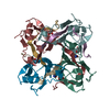
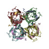
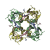
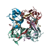
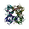
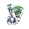
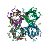
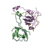
- PDB-7a8y: X-ray crystal structure of Aspartate alpha-decarboxylase in compl... -
+ Open data

- Basic information
| Entry | Database: PDB / ID: 7a8y | ||||||||||||
|---|---|---|---|---|---|---|---|---|---|---|---|---|---|
| Title | X-ray crystal structure of Aspartate alpha-decarboxylase in complex with D-Serine | ||||||||||||
 Components Components | (Aspartate 1-decarboxylase) x 2 | ||||||||||||
 Keywords Keywords | LYASE / Decarboxylase | ||||||||||||
| Function / homology |  Function and homology information Function and homology informationalanine biosynthetic process / aspartate 1-decarboxylase / aspartate 1-decarboxylase activity / pantothenate biosynthetic process / cytoplasm Similarity search - Function | ||||||||||||
| Biological species |  | ||||||||||||
| Method |  X-RAY DIFFRACTION / SYNCHROTRON / MOLECULAR REPLACEMENT / Resolution: 1.75 Å X-RAY DIFFRACTION / SYNCHROTRON / MOLECULAR REPLACEMENT / Resolution: 1.75 Å | ||||||||||||
 Authors Authors | Yorke, B.A. / Raskar, T. | ||||||||||||
| Funding support |  United Kingdom, 1items United Kingdom, 1items
| ||||||||||||
 Citation Citation | Journal: Phys Chem Chem Phys / Year: 2022 Title: Structure and diffusive dynamics of aspartate alpha-decarboxylase (ADC) liganded with D-serine in aqueous solution. Authors: Raskar, T. / Niebling, S. / Devos, J.M. / Yorke, B.A. / Hartlein, M. / Huse, N. / Forsyth, V.T. / Seydel, T. / Pearson, A.R. | ||||||||||||
| History |
|
- Structure visualization
| Structure viewer | Molecule: MolmilJmol/JSmol |
|---|
- Downloads & links
-Download
| PDBx/mmCIF format | 7a8y.cif.gz | 72 KB | Display | PDBx/mmCIF format |
|---|---|---|---|---|
| PDB format | pdb7a8y.ent.gz | Display | PDB format | |
| PDBx/mmJSON format | 7a8y.json.gz | Tree view | PDBx/mmJSON format | |
| Others |  Other downloads Other downloads |
-Validation report
| Arichive directory | https://data.pdbj.org/pub/pdb/validation_reports/a8/7a8yftp://data.pdbj.org/pub/pdb/validation_reports/a8/7a8y | HTTPS FTP |
|---|
-Related structure data
| Related structure data |  1aw8S S: Starting model for refinement |
|---|---|
| Similar structure data |


-Links
 PDBj
PDBj


- Assembly
| Deposited unit | 
| ||||||||
|---|---|---|---|---|---|---|---|---|---|
| 1 | 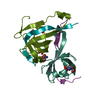
| ||||||||
| Unit cell |
|
-Components
-Protein/peptide / Protein , 2 types, 4 molecules AAADDDBBBEaE
| #1: Protein/peptide | Mass: 3056.588 Da / Num. of mol.: 2 Source method: isolated from a genetically manipulated source Details: Modification at residue 25: Serine and pyrovoyl residues are present with partial occupancies. Source: (gene. exp.) Gene: panD, BON96_17415, D9J78_12565, FKO60_12525, G5697_15675 Production host: #2: Protein | Mass: 10219.350 Da / Num. of mol.: 2 Source method: isolated from a genetically manipulated source Details: Some of the terminal residues are not modeled due to missing density Source: (gene. exp.) Gene: panD, BON96_17415, D9J78_12565, FKO60_12525, G5697_15675 Production host: |
|---|
-Non-polymers , 4 types, 213 molecules 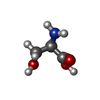


| #3: Chemical |  Type: D-peptide linking / Mass: 105.093 Da / Num. of mol.: 3 / Source method: obtained synthetically / Formula: C3H7NO3 / Feature type: SUBJECT OF INVESTIGATION Type: D-peptide linking / Mass: 105.093 Da / Num. of mol.: 3 / Source method: obtained synthetically / Formula: C3H7NO3 / Feature type: SUBJECT OF INVESTIGATION#4: Chemical | ChemComp-EDO / |  Mass: 62.068 Da / Num. of mol.: 1 / Source method: obtained synthetically / Formula: C2H6O2 Mass: 62.068 Da / Num. of mol.: 1 / Source method: obtained synthetically / Formula: C2H6O2#5: Chemical | ChemComp-SER / |  Type: L-peptide linking / Mass: 105.093 Da / Num. of mol.: 1 / Source method: obtained synthetically / Formula: C3H7NO3 Type: L-peptide linking / Mass: 105.093 Da / Num. of mol.: 1 / Source method: obtained synthetically / Formula: C3H7NO3#6: Water | ChemComp-HOH / | Mass: 18.015 Da / Num. of mol.: 208 / Source method: isolated from a natural source / Formula: H2O |
|---|
-Details
| Has ligand of interest | Y |
|---|---|
| Has protein modification | Y |
-Experimental details
-Experiment
| Experiment | Method: X-RAY DIFFRACTION / Number of used crystals: 1 |
|---|
- Sample preparation
| Crystal | Density Matthews: 3.11 Å3/Da / Density % sol: 60.49 % |
|---|---|
| Crystal grow | Temperature: 293 K / Method: vapor diffusion / pH: 4.5 Details: 1.8 M ammonium sulphate, 100 mM sodium citrate pH 4.5 |
-Data collection
| Diffraction | Mean temperature: 100 K / Serial crystal experiment: N |
|---|---|
| Diffraction source | Source: SYNCHROTRON / Site: Diamond / Beamline: I24 / Wavelength: 0.9778 Å |
| Detector | Type: DECTRIS PILATUS 6M / Detector: PIXEL / Date: Nov 23, 2010 |
| Radiation | Protocol: SINGLE WAVELENGTH / Monochromatic (M) / Laue (L): M / Scattering type: x-ray |
| Radiation wavelength | Wavelength: 0.9778 Å / Relative weight: 1 |
| Reflection | Resolution: 1.75→46.98 Å / Num. obs: 33922 / % possible obs: 99.99 % / Redundancy: 2 % / CC1/2: 0.99 / Rmerge(I) obs: 0.02384 / Net I/σ(I): 21.5 |
| Reflection shell | Resolution: 1.75→1.81 Å / Rmerge(I) obs: 0.1564 / Mean I/σ(I) obs: 5.25 / Num. unique obs: 3301 / CC1/2: 0.93 |
- Processing
| Software |
| |||||||||||||||||||||||||||||||||||||||||||||||||||||||||||||||||||||||||||||||||||||||||||||||||||||||||||||||||||||||||||||||||||||||||||||||||||||||||||
|---|---|---|---|---|---|---|---|---|---|---|---|---|---|---|---|---|---|---|---|---|---|---|---|---|---|---|---|---|---|---|---|---|---|---|---|---|---|---|---|---|---|---|---|---|---|---|---|---|---|---|---|---|---|---|---|---|---|---|---|---|---|---|---|---|---|---|---|---|---|---|---|---|---|---|---|---|---|---|---|---|---|---|---|---|---|---|---|---|---|---|---|---|---|---|---|---|---|---|---|---|---|---|---|---|---|---|---|---|---|---|---|---|---|---|---|---|---|---|---|---|---|---|---|---|---|---|---|---|---|---|---|---|---|---|---|---|---|---|---|---|---|---|---|---|---|---|---|---|---|---|---|---|---|---|---|---|
| Refinement | Method to determine structure: MOLECULAR REPLACEMENT Starting model: 1AW8 Resolution: 1.75→46.975 Å / Cor.coef. Fo:Fc: 0.961 / Cor.coef. Fo:Fc free: 0.947 / WRfactor Rfree: 0.187 / WRfactor Rwork: 0.165 / SU B: 1.67 / SU ML: 0.054 / Average fsc free: 0.9557 / Average fsc work: 0.9597 / Cross valid method: FREE R-VALUE / ESU R: 0.092 / ESU R Free: 0.088 Details: Hydrogens have been added in their riding positions
| |||||||||||||||||||||||||||||||||||||||||||||||||||||||||||||||||||||||||||||||||||||||||||||||||||||||||||||||||||||||||||||||||||||||||||||||||||||||||||
| Solvent computation | Ion probe radii: 0.8 Å / Shrinkage radii: 0.8 Å / VDW probe radii: 1.2 Å / Solvent model: MASK BULK SOLVENT | |||||||||||||||||||||||||||||||||||||||||||||||||||||||||||||||||||||||||||||||||||||||||||||||||||||||||||||||||||||||||||||||||||||||||||||||||||||||||||
| Displacement parameters | Biso mean: 20.673 Å2
| |||||||||||||||||||||||||||||||||||||||||||||||||||||||||||||||||||||||||||||||||||||||||||||||||||||||||||||||||||||||||||||||||||||||||||||||||||||||||||
| Refinement step | Cycle: LAST / Resolution: 1.75→46.975 Å
| |||||||||||||||||||||||||||||||||||||||||||||||||||||||||||||||||||||||||||||||||||||||||||||||||||||||||||||||||||||||||||||||||||||||||||||||||||||||||||
| Refine LS restraints |
| |||||||||||||||||||||||||||||||||||||||||||||||||||||||||||||||||||||||||||||||||||||||||||||||||||||||||||||||||||||||||||||||||||||||||||||||||||||||||||
| LS refinement shell |
|
