 ムービー
ムービー コントローラー


+ データを開く

- 基本情報
基本情報
| 登録情報 | データベース: PDB / ID: 7a7d | ||||||
|---|---|---|---|---|---|---|---|
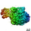
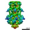
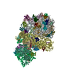
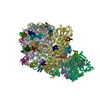
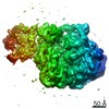
| タイトル | Cadherin fit into cryo-ET map | ||||||
 要素 要素 |
| ||||||
 キーワード キーワード | CELL ADHESION / Cadherin / Desmosome | ||||||
| 機能・相同性 |  機能・相同性情報 機能・相同性情報cardiac muscle cell-cardiac muscle cell adhesion / Purkinje myocyte development / positive regulation of protein localization to cell-cell junction / bundle of His cell-Purkinje myocyte adhesion involved in cell communication / cell adhesive protein binding involved in bundle of His cell-Purkinje myocyte communication / desmosome organization / negative regulation of endothelial cell differentiation / Keratinization / negative regulation of inflammatory response to wounding / desmosome ...cardiac muscle cell-cardiac muscle cell adhesion / Purkinje myocyte development / positive regulation of protein localization to cell-cell junction / bundle of His cell-Purkinje myocyte adhesion involved in cell communication / cell adhesive protein binding involved in bundle of His cell-Purkinje myocyte communication / desmosome organization / negative regulation of endothelial cell differentiation / Keratinization / negative regulation of inflammatory response to wounding / desmosome / mesenchymal to epithelial transition / Formation of the cornified envelope / cornified envelope / regulation of ventricular cardiac muscle cell action potential / Apoptotic cleavage of cell adhesion proteins / positive regulation of p38MAPK cascade / negative regulation of epithelial to mesenchymal transition / positive regulation of sprouting angiogenesis / homophilic cell-cell adhesion / positive regulation of stem cell population maintenance / regulation of heart rate by cardiac conduction / intercalated disc / RHOG GTPase cycle / lateral plasma membrane / RAC2 GTPase cycle / maternal process involved in female pregnancy / RAC3 GTPase cycle / cell adhesion molecule binding / positive regulation of cell adhesion / response to progesterone / cellular response to starvation / stem cell proliferation / adherens junction / cell-cell adhesion / cell-cell junction / cell junction / cytoplasmic vesicle / cell adhesion / apical plasma membrane / intracellular membrane-bounded organelle / calcium ion binding / negative regulation of apoptotic process / cell surface / endoplasmic reticulum / extracellular exosome / plasma membrane / cytoplasm 類似検索 - 分子機能 | ||||||
| 生物種 |  Homo sapiens (ヒト) Homo sapiens (ヒト) | ||||||
| 手法 | 電子顕微鏡法 / サブトモグラム平均法 / クライオ電子顕微鏡法 / 解像度: 26 Å | ||||||
 データ登録者 データ登録者 | Sikora, M. / Ermel, U.H. / Seybold, A. / Kunz, M. / Calloni, G. / Reitz, J. / Vabulas, R.M. / Hummer, G. / Frangakis, A.S. | ||||||
 引用 引用 | ジャーナル: Proc Natl Acad Sci U S A / 年: 2020 タイトル: Desmosome architecture derived from molecular dynamics simulations and cryo-electron tomography. 著者: Mateusz Sikora / Utz H Ermel / Anna Seybold / Michael Kunz / Giulia Calloni / Julian Reitz / R Martin Vabulas / Gerhard Hummer / Achilleas S Frangakis /   要旨: Desmosomes are cell-cell junctions that link tissue cells experiencing intense mechanical stress. Although the structure of the desmosomal cadherins is known, the desmosome architecture-which is ...Desmosomes are cell-cell junctions that link tissue cells experiencing intense mechanical stress. Although the structure of the desmosomal cadherins is known, the desmosome architecture-which is essential for mediating numerous functions-remains elusive. Here, we recorded cryo-electron tomograms (cryo-ET) in which individual cadherins can be discerned; they appear variable in shape, spacing, and tilt with respect to the membrane. The resulting sub-tomogram average reaches a resolution of ∼26 Å, limited by the inherent flexibility of desmosomes. To address this challenge typical of dynamic biological assemblies, we combine sub-tomogram averaging with atomistic molecular dynamics (MD) simulations. We generate models of possible cadherin arrangements and perform an in silico screening according to biophysical and structural properties extracted from MD simulation trajectories. We find a truss-like arrangement of cadherins that resembles the characteristic footprint seen in the electron micrograph. The resulting model of the desmosomal architecture explains their unique biophysical properties and strength. | ||||||
| 履歴 |
|
- 構造の表示
構造の表示
| ムービー |
ムービービューア |
|---|---|
| 構造ビューア | 分子: MolmilJmol/JSmol |
- ダウンロードとリンク
-ダウンロード
| PDBx/mmCIF形式 | 7a7d.cif.gz | 2.2 MB | 表示 | PDBx/mmCIF形式 |
|---|---|---|---|---|
| PDB形式 | pdb7a7d.ent.gz | 表示 | PDB形式 | |
| PDBx/mmJSON形式 | 7a7d.json.gz | ツリー表示 | PDBx/mmJSON形式 | |
| その他 |  その他のダウンロード その他のダウンロード |
-検証レポート
| 文書・要旨 | 7a7d_validation.pdf.gz | 879.3 KB | 表示 | wwPDB検証レポート |
|---|---|---|---|---|
| 文書・詳細版 | 7a7d_full_validation.pdf.gz | 1.1 MB | 表示 | |
| XML形式データ | 7a7d_validation.xml.gz | 194.7 KB | 表示 | |
| CIF形式データ | 7a7d_validation.cif.gz | 301.7 KB | 表示 | |
| アーカイブディレクトリ | https://data.pdbj.org/pub/pdb/validation_reports/a7/7a7dftp://data.pdbj.org/pub/pdb/validation_reports/a7/7a7d | HTTPS FTP |
-関連構造データ


-リンク
 PDBj
PDBj


- 集合体
集合体
| 登録構造単位 | 
|
|---|---|
| 1 |
|
-要素
| #1: タンパク質 | 分子量: 61893.297 Da / 分子数: 7 / 由来タイプ: 天然 / 由来: (天然) Homo sapiens (ヒト) / 参照: UniProt: Q14126#2: タンパク質 | 分子量: 60869.840 Da / 分子数: 7 / 由来タイプ: 天然 / 由来: (天然) Homo sapiens (ヒト) / 参照: UniProt: Q02487研究の焦点であるリガンドがあるか | N | Has protein modification | Y | |
|---|
-実験情報
-実験
| 実験 | 手法: 電子顕微鏡法 |
|---|---|
| EM実験 | 試料の集合状態: TISSUE / 3次元再構成法: サブトモグラム平均法 |
- 試料調製
試料調製
| 構成要素 | 名称: Desmosome from mouse liver / タイプ: ORGANELLE OR CELLULAR COMPONENT / Entity ID: all / 由来: NATURAL |
|---|---|
| 由来(天然) | 生物種: Homo sapiens (ヒト) |
| 緩衝液 | pH: 7.5 |
| 試料 | 包埋: NO / シャドウイング: NO / 染色: NO / 凍結: YES |
| 急速凍結 | 凍結剤: ETHANE |
- 電子顕微鏡撮影
| 実験機器 |  モデル: Titan Krios / 画像提供: FEI Company |
|---|---|
| 顕微鏡 | モデル: FEI TITAN KRIOS |
| 電子銃 | 電子線源:  FIELD EMISSION GUN / 加速電圧: 300 kV / 照射モード: FLOOD BEAM FIELD EMISSION GUN / 加速電圧: 300 kV / 照射モード: FLOOD BEAM |
| 電子レンズ | モード: BRIGHT FIELD / 倍率(公称値): 64000 X / Cs: 2.7 mm |
| 試料ホルダ | 凍結剤: NITROGEN 試料ホルダーモデル: FEI TITAN KRIOS AUTOGRID HOLDER |
| 撮影 | 平均露光時間: 2 sec. / 電子線照射量: 1.95 e/Å2 / 検出モード: SUPER-RESOLUTION フィルム・検出器のモデル: GATAN K2 SUMMIT (4k x 4k) |
| 電子光学装置 | エネルギーフィルター名称: GIF Quantum SE |
| 画像スキャン | 動画フレーム数/画像: 4 |
- 解析
解析
| EMソフトウェア | 名称: SerialEM / カテゴリ: 画像取得 |
|---|---|
| CTF補正 | タイプ: PHASE FLIPPING AND AMPLITUDE CORRECTION |
| 対称性 | 点対称性: C1 (非対称) |
| 3次元再構成 | 解像度: 26 Å / 解像度の算出法: FSC 0.5 CUT-OFF / 粒子像の数: 3656 / 対称性のタイプ: POINT |
| EM volume selection | Num. of tomograms: 12 / Num. of volumes extracted: 9000 |
