 Movie
Movie Controller

[English] 日本語
 Yorodumi
- PDB-7a4x: tRNA-guanine transglycosylase C158S/C281S/Y330C/H333A mutant in c... -
+ Open data

- Basic information
| Entry | Database: PDB / ID: 7a4x | ||||||
|---|---|---|---|---|---|---|---|
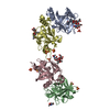
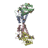
| Title | tRNA-guanine transglycosylase C158S/C281S/Y330C/H333A mutant in complex with dimethyl sulfoxide | ||||||
 Components Components | Queuine tRNA-ribosyltransferase | ||||||
 Keywords Keywords | TRANSFERASE / enzyme / disulfide / fragment | ||||||
| Function / homology |  Function and homology information Function and homology informationtRNA-guanosine34 preQ1 transglycosylase / tRNA-guanosine(34) queuine transglycosylase activity / tRNA queuosine(34) biosynthetic process / metal ion binding / cytosol Similarity search - Function | ||||||
| Biological species |  Zymomonas mobilis subsp. mobilis ZM4 = ATCC 31821 (bacteria) Zymomonas mobilis subsp. mobilis ZM4 = ATCC 31821 (bacteria) | ||||||
| Method |  X-RAY DIFFRACTION / SYNCHROTRON / MOLECULAR REPLACEMENT / Resolution: 2.05 Å X-RAY DIFFRACTION / SYNCHROTRON / MOLECULAR REPLACEMENT / Resolution: 2.05 Å | ||||||
 Authors Authors | Nguyen, D. / Nguyen, T.X.P. / Heine, A. / Klebe, G. | ||||||
| Funding support |  Germany, 1items Germany, 1items
| ||||||
 Citation Citation | Journal: Acs Chem.Biol. / Year: 2021 Title: Targeting a Cryptic Pocket in a Protein-Protein Contact by Disulfide-Induced Rupture of a Homodimeric Interface. Authors: Nguyen, D. / Xie, X. / Jakobi, S. / Terwesten, F. / Metz, A. / Nguyen, T.X.P. / Palchykov, V.A. / Heine, A. / Reuter, K. / Klebe, G. | ||||||
| History |
|
- Structure visualization
| Structure viewer | Molecule: MolmilJmol/JSmol |
|---|
- Downloads & links
-Download
| PDBx/mmCIF format | 7a4x.cif.gz | 198.1 KB | Display | PDBx/mmCIF format |
|---|---|---|---|---|
| PDB format | pdb7a4x.ent.gz | 130.3 KB | Display | PDB format |
| PDBx/mmJSON format | 7a4x.json.gz | Tree view | PDBx/mmJSON format | |
| Others |  Other downloads Other downloads |
-Validation report
| Arichive directory | https://data.pdbj.org/pub/pdb/validation_reports/a4/7a4xftp://data.pdbj.org/pub/pdb/validation_reports/a4/7a4x | HTTPS FTP |
|---|
-Related structure data
| Related structure data |  4jbrC  7a0bC  7a3vC  7a3xC  7a4kC  7a6dC  7a9eC  7adnC  1pudS C: citing same article ( S: Starting model for refinement |
|---|---|
| Similar structure data |


-Links
 PDBj
PDBj- Assembly
| Deposited unit | 
| ||||||||||||
|---|---|---|---|---|---|---|---|---|---|---|---|---|---|
| 1 | 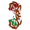
| ||||||||||||
| Unit cell |
| ||||||||||||
| Components on special symmetry positions |
|
-Components
-Protein , 1 types, 1 molecules A
| #1: Protein | Mass: 42910.605 Da / Num. of mol.: 1 / Mutation: C158S/C281S/Y330C/H333A Source method: isolated from a genetically manipulated source Source: (gene. exp.) Zymomonas mobilis subsp. mobilis ZM4 = ATCC 31821 (bacteria)Gene: tgt, ZMO0363 / Plasmid: pPR-IBA2 / Production host: References: UniProt: P28720, tRNA-guanosine34 preQ1 transglycosylase |
|---|
-Non-polymers , 5 types, 188 molecules 


| #2: Chemical | ChemComp-ZN /  Mass: 65.409 Da / Num. of mol.: 1 / Source method: obtained synthetically / Formula: Zn Mass: 65.409 Da / Num. of mol.: 1 / Source method: obtained synthetically / Formula: Zn | ||||||
|---|---|---|---|---|---|---|---|
| #3: Chemical |  Mass: 92.094 Da / Num. of mol.: 2 / Source method: obtained synthetically / Formula: C3H8O3 Mass: 92.094 Da / Num. of mol.: 2 / Source method: obtained synthetically / Formula: C3H8O3#4: Chemical | ChemComp-DMS / |  Mass: 78.133 Da / Num. of mol.: 1 / Source method: obtained synthetically / Formula: C2H6OS / Feature type: SUBJECT OF INVESTIGATION / Comment: DMSO, precipitant*YM Mass: 78.133 Da / Num. of mol.: 1 / Source method: obtained synthetically / Formula: C2H6OS / Feature type: SUBJECT OF INVESTIGATION / Comment: DMSO, precipitant*YM#5: Chemical | ChemComp-EDO / |  Mass: 62.068 Da / Num. of mol.: 1 / Source method: obtained synthetically / Formula: C2H6O2 Mass: 62.068 Da / Num. of mol.: 1 / Source method: obtained synthetically / Formula: C2H6O2#6: Water | ChemComp-HOH / | Mass: 18.015 Da / Num. of mol.: 183 / Source method: isolated from a natural source / Formula: H2O |
-Details
| Has ligand of interest | Y |
|---|---|
| Has protein modification | Y |
-Experimental details
-Experiment
| Experiment | Method: X-RAY DIFFRACTION / Number of used crystals: 1 |
|---|
- Sample preparation
| Crystal | Density Matthews: 3.16 Å3/Da / Density % sol: 61.06 % |
|---|---|
| Crystal grow | Temperature: 291.15 K / Method: vapor diffusion, sitting drop / pH: 8.5 / Details: 0.1 M TRIS, pH 8.5, 5% PEG 6000 |
-Data collection
| Diffraction | Mean temperature: 100 K / Serial crystal experiment: N |
|---|---|
| Diffraction source | Source: SYNCHROTRON / Site: PETRA III, EMBL c/o DESY / Beamline: P13 (MX1) / Wavelength: 0.7999 Å |
| Detector | Type: DECTRIS PILATUS 6M-F / Detector: PIXEL / Date: Nov 6, 2015 |
| Radiation | Protocol: SINGLE WAVELENGTH / Monochromatic (M) / Laue (L): M / Scattering type: x-ray |
| Radiation wavelength | Wavelength: 0.7999 Å / Relative weight: 1 |
| Reflection | Resolution: 2.05→64.06 Å / Num. obs: 35368 / % possible obs: 99.9 % / Redundancy: 21.9 % / Biso Wilson estimate: 31.67 Å2 / CC1/2: 0.999 / Rsym value: 0.1 / Net I/σ(I): 22.3 |
| Reflection shell | Resolution: 2.05→2.17 Å / Mean I/σ(I) obs: 5.7 / Num. unique obs: 5626 / CC1/2: 0.957 / Rsym value: 0.508 / % possible all: 99.3 |
- Processing
| Software |
| |||||||||||||||||||||||||||||||||||||||||||||||||||||||||||||||||||||||||||||||||||||||||||
|---|---|---|---|---|---|---|---|---|---|---|---|---|---|---|---|---|---|---|---|---|---|---|---|---|---|---|---|---|---|---|---|---|---|---|---|---|---|---|---|---|---|---|---|---|---|---|---|---|---|---|---|---|---|---|---|---|---|---|---|---|---|---|---|---|---|---|---|---|---|---|---|---|---|---|---|---|---|---|---|---|---|---|---|---|---|---|---|---|---|---|---|---|
| Refinement | Method to determine structure: MOLECULAR REPLACEMENT Starting model: 1PUD Resolution: 2.05→64.06 Å / SU ML: 0.1774 / Cross valid method: FREE R-VALUE / σ(F): 1.38 / Phase error: 18.3551 Stereochemistry target values: GeoStd + Monomer Library + CDL v1.2
| |||||||||||||||||||||||||||||||||||||||||||||||||||||||||||||||||||||||||||||||||||||||||||
| Solvent computation | Shrinkage radii: 0.9 Å / VDW probe radii: 1.11 Å / Solvent model: FLAT BULK SOLVENT MODEL | |||||||||||||||||||||||||||||||||||||||||||||||||||||||||||||||||||||||||||||||||||||||||||
| Displacement parameters | Biso mean: 38.69 Å2 | |||||||||||||||||||||||||||||||||||||||||||||||||||||||||||||||||||||||||||||||||||||||||||
| Refinement step | Cycle: LAST / Resolution: 2.05→64.06 Å
| |||||||||||||||||||||||||||||||||||||||||||||||||||||||||||||||||||||||||||||||||||||||||||
| Refine LS restraints |
| |||||||||||||||||||||||||||||||||||||||||||||||||||||||||||||||||||||||||||||||||||||||||||
| LS refinement shell |
| |||||||||||||||||||||||||||||||||||||||||||||||||||||||||||||||||||||||||||||||||||||||||||
| Refinement TLS params. | Method: refined / Refine-ID: X-RAY DIFFRACTION
| |||||||||||||||||||||||||||||||||||||||||||||||||||||||||||||||||||||||||||||||||||||||||||
| Refinement TLS group |
|
