 Movie
Movie Controller

[English] 日本語
 Yorodumi
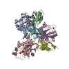
- PDB-7a0v: Crystal structure of the 5-phosphatase domain of Synaptojanin1 in... -
+ Open data

- Basic information
| Entry | Database: PDB / ID: 7a0v | ||||||||||||
|---|---|---|---|---|---|---|---|---|---|---|---|---|---|
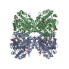
| Title | Crystal structure of the 5-phosphatase domain of Synaptojanin1 in complex with a nanobody | ||||||||||||
 Components Components |
| ||||||||||||
 Keywords Keywords | HYDROLASE / Inositol polyphosphate 5-phosphatase / Phosphoinositide / Parkinson's disease / Epilepsy | ||||||||||||
| Function / homology |  Function and homology information Function and homology informationpositive regulation of endosome organization / phosphatidylinositol phosphate 5-phosphatase activity / phosphatidylinositol-4-phosphate phosphatase activity / phosphatidylinositol phosphate 4-phosphatase activity / phosphatidylinositol-3,5-bisphosphate 5-phosphatase activity / phosphatidylinositol-3,5-bisphosphate 3-phosphatase activity / inositol-1,4,5-trisphosphate 5-phosphatase activity / phosphatidylinositol-3-phosphate phosphatase activity / Synthesis of IP2, IP, and Ins in the cytosol / synaptic vesicle uncoating ...positive regulation of endosome organization / phosphatidylinositol phosphate 5-phosphatase activity / phosphatidylinositol-4-phosphate phosphatase activity / phosphatidylinositol phosphate 4-phosphatase activity / phosphatidylinositol-3,5-bisphosphate 5-phosphatase activity / phosphatidylinositol-3,5-bisphosphate 3-phosphatase activity / inositol-1,4,5-trisphosphate 5-phosphatase activity / phosphatidylinositol-3-phosphate phosphatase activity / Synthesis of IP2, IP, and Ins in the cytosol / synaptic vesicle uncoating / inositol phosphate metabolic process / clathrin coat of coated pit / phosphoinositide 5-phosphatase / phosphatidylinositol-4,5-bisphosphate 5-phosphatase activity / phosphatidylinositol metabolic process / phosphatidylinositol dephosphorylation / membrane coat / phosphatidylinositol biosynthetic process / membrane organization / vesicle membrane / neurotransmitter transport / synaptic vesicle priming / Synthesis of IP3 and IP4 in the cytosol / Synthesis of PIPs at the plasma membrane / synaptic vesicle transport / synaptic vesicle endocytosis / learning / synaptic membrane / terminal bouton / Clathrin-mediated endocytosis / presynapse / perinuclear region of cytoplasm / RNA binding / membrane / cytoplasm / cytosol Similarity search - Function | ||||||||||||
| Biological species |  Homo sapiens (human) Homo sapiens (human) | ||||||||||||
| Method |  X-RAY DIFFRACTION / SYNCHROTRON / MOLECULAR REPLACEMENT / Resolution: 2.3 Å X-RAY DIFFRACTION / SYNCHROTRON / MOLECULAR REPLACEMENT / Resolution: 2.3 Å | ||||||||||||
 Authors Authors | Paesmans, J. / Galicia, C. / Martin, E. / Versees, W. | ||||||||||||
| Funding support |  Belgium, 3items Belgium, 3items
| ||||||||||||
 Citation Citation | Journal: Elife / Year: 2020 Title: A structure of substrate-bound Synaptojanin1 provides new insights in its mechanism and the effect of disease mutations. Authors: Paesmans, J. / Martin, E. / Deckers, B. / Berghmans, M. / Sethi, R. / Loeys, Y. / Pardon, E. / Steyaert, J. / Verstreken, P. / Galicia, C. / Versees, W. | ||||||||||||
| History |
|
- Structure visualization
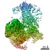
| Structure viewer | Molecule: MolmilJmol/JSmol |
|---|
- Downloads & links
-Download
| PDBx/mmCIF format | 7a0v.cif.gz | 287.7 KB | Display | PDBx/mmCIF format |
|---|---|---|---|---|
| PDB format | pdb7a0v.ent.gz | 226.4 KB | Display | PDB format |
| PDBx/mmJSON format | 7a0v.json.gz | Tree view | PDBx/mmJSON format | |
| Others |  Other downloads Other downloads |
-Validation report
| Arichive directory | https://data.pdbj.org/pub/pdb/validation_reports/a0/7a0vftp://data.pdbj.org/pub/pdb/validation_reports/a0/7a0v | HTTPS FTP |
|---|
-Related structure data
| Related structure data |  7a17C  1i9yS 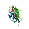 3mtcS 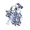 3n9vS 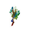 4cmnS  4nc2S S: Starting model for refinement C: citing same article ( |
|---|---|
| Similar structure data |
-Links
 PDBj
PDBj


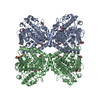
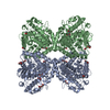
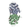
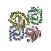
- Assembly
| Deposited unit | 
| ||||||||||||
|---|---|---|---|---|---|---|---|---|---|---|---|---|---|
| 1 |
| ||||||||||||
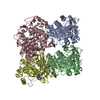
| Unit cell |
| ||||||||||||
| Components on special symmetry positions |
|
-Components
-Protein / Antibody , 2 types, 6 molecules ACEBDF
| #1: Protein | Mass: 39866.387 Da / Num. of mol.: 3 Source method: isolated from a genetically manipulated source Source: (gene. exp.) Homo sapiens (human) / Gene: SYNJ1, KIAA0910 / Plasmid: pET28a / Production host:  #2: Antibody | Mass: 14559.901 Da / Num. of mol.: 3 Source method: isolated from a genetically manipulated source Source: (gene. exp.) |
|---|
-Non-polymers , 4 types, 369 molecules 


| #3: Chemical |  Mass: 94.971 Da / Num. of mol.: 3 / Source method: obtained synthetically / Formula: PO4 Mass: 94.971 Da / Num. of mol.: 3 / Source method: obtained synthetically / Formula: PO4#4: Chemical |  Mass: 24.305 Da / Num. of mol.: 3 / Source method: obtained synthetically / Formula: Mg Mass: 24.305 Da / Num. of mol.: 3 / Source method: obtained synthetically / Formula: Mg#5: Chemical | ChemComp-GOL /  Mass: 92.094 Da / Num. of mol.: 4 / Source method: obtained synthetically / Formula: C3H8O3 Mass: 92.094 Da / Num. of mol.: 4 / Source method: obtained synthetically / Formula: C3H8O3#6: Water | ChemComp-HOH / | Mass: 18.015 Da / Num. of mol.: 359 / Source method: isolated from a natural source / Formula: H2O |
|---|
-Details
| Has ligand of interest | N |
|---|---|
| Has protein modification | Y |
-Experimental details
-Experiment
| Experiment | Method: X-RAY DIFFRACTION / Number of used crystals: 1 |
|---|
- Sample preparation
| Crystal | Density Matthews: 2.61 Å3/Da / Density % sol: 52.83 % |
|---|---|
| Crystal grow | Temperature: 293 K / Method: vapor diffusion, sitting drop / pH: 5 Details: 15% PEG 4000, 0.1 M sodium citrate pH 5, 10% 2-propanol |
-Data collection
| Diffraction | Mean temperature: 100 K / Serial crystal experiment: N |
|---|---|
| Diffraction source | Source: SYNCHROTRON / Site: Diamond  / Beamline: I03 / Wavelength: 0.980105 Å / Beamline: I03 / Wavelength: 0.980105 Å |
| Detector | Type: DECTRIS EIGER2 XE 16M / Detector: PIXEL / Date: Nov 27, 2019 |
| Radiation | Protocol: SINGLE WAVELENGTH / Monochromatic (M) / Laue (L): M / Scattering type: x-ray |
| Radiation wavelength | Wavelength: 0.980105 Å / Relative weight: 1 |
| Reflection | Resolution: 2.297→86.81 Å / Num. obs: 53823 / % possible obs: 92.3 % / Redundancy: 7 % / Biso Wilson estimate: 38.08 Å2 / CC1/2: 0.997 / Rmerge(I) obs: 0.133 / Rpim(I) all: 0.054 / Rrim(I) all: 0.143 / Net I/σ(I): 11.2 |
| Reflection shell | Resolution: 2.3→2.43 Å / Num. unique obs: 1511 / CC1/2: 0.512 |
- Processing
| Software |
| |||||||||||||||||||||||||||||||||||||||||||||||||||||||||||||||||||||||||||||||||||||||||||||||||||||||||||||||||||||||||||||||||||||||||||||||||||
|---|---|---|---|---|---|---|---|---|---|---|---|---|---|---|---|---|---|---|---|---|---|---|---|---|---|---|---|---|---|---|---|---|---|---|---|---|---|---|---|---|---|---|---|---|---|---|---|---|---|---|---|---|---|---|---|---|---|---|---|---|---|---|---|---|---|---|---|---|---|---|---|---|---|---|---|---|---|---|---|---|---|---|---|---|---|---|---|---|---|---|---|---|---|---|---|---|---|---|---|---|---|---|---|---|---|---|---|---|---|---|---|---|---|---|---|---|---|---|---|---|---|---|---|---|---|---|---|---|---|---|---|---|---|---|---|---|---|---|---|---|---|---|---|---|---|---|---|---|
| Refinement | Method to determine structure: MOLECULAR REPLACEMENT Starting model: 1i9y, 3n9v, 3mtc, 4cmn, 4nc2 Resolution: 2.3→86.81 Å / SU ML: 0.3193 / Cross valid method: FREE R-VALUE / σ(F): 1.34 / Phase error: 29.8898 Stereochemistry target values: GeoStd + Monomer Library + CDL v1.2
| |||||||||||||||||||||||||||||||||||||||||||||||||||||||||||||||||||||||||||||||||||||||||||||||||||||||||||||||||||||||||||||||||||||||||||||||||||
| Solvent computation | Shrinkage radii: 0.9 Å / VDW probe radii: 1.11 Å / Solvent model: FLAT BULK SOLVENT MODEL | |||||||||||||||||||||||||||||||||||||||||||||||||||||||||||||||||||||||||||||||||||||||||||||||||||||||||||||||||||||||||||||||||||||||||||||||||||
| Displacement parameters | Biso mean: 47.27 Å2 | |||||||||||||||||||||||||||||||||||||||||||||||||||||||||||||||||||||||||||||||||||||||||||||||||||||||||||||||||||||||||||||||||||||||||||||||||||
| Refinement step | Cycle: LAST / Resolution: 2.3→86.81 Å
| |||||||||||||||||||||||||||||||||||||||||||||||||||||||||||||||||||||||||||||||||||||||||||||||||||||||||||||||||||||||||||||||||||||||||||||||||||
| Refine LS restraints |
| |||||||||||||||||||||||||||||||||||||||||||||||||||||||||||||||||||||||||||||||||||||||||||||||||||||||||||||||||||||||||||||||||||||||||||||||||||
| LS refinement shell |
|
