 Movie
Movie Controller


+ Open data

- Basic information
| Entry | Database: PDB / ID: 6zxu | ||||||
|---|---|---|---|---|---|---|---|
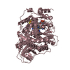
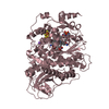
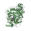
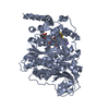
| Title | Catabolic reductive dehalogenase NpRdhA, N-terminally tagged. | ||||||
 Components Components | Oxidoreductase, NAD-binding/iron-sulfur cluster-binding protein | ||||||
 Keywords Keywords | OXIDOREDUCTASE / reductive dehalogenase / B12 / iron-sulfur | ||||||
| Function / homology |  Function and homology information Function and homology information | ||||||
| Biological species |  Nitratireductor pacificus pht-3B (bacteria) Nitratireductor pacificus pht-3B (bacteria) | ||||||
| Method |  X-RAY DIFFRACTION / SYNCHROTRON / MOLECULAR REPLACEMENT / Resolution: 1.73 Å X-RAY DIFFRACTION / SYNCHROTRON / MOLECULAR REPLACEMENT / Resolution: 1.73 Å | ||||||
 Authors Authors | Leys, D. / Halliwell, T. | ||||||
| Funding support |  United Kingdom, 1items United Kingdom, 1items
| ||||||
 Citation Citation | Journal: Microorganisms / Year: 2020 Title: Catabolic Reductive Dehalogenase Substrate Complex Structures Underpin Rational Repurposing of Substrate Scope. Authors: Halliwell, T. / Fisher, K. / Payne, K.A.P. / Rigby, S.E.J. / Leys, D. | ||||||
| History |
|
- Structure visualization
| Structure viewer | Molecule: MolmilJmol/JSmol |
|---|
- Downloads & links
-Download
| PDBx/mmCIF format | 6zxu.cif.gz | 452.3 KB | Display | PDBx/mmCIF format |
|---|---|---|---|---|
| PDB format | pdb6zxu.ent.gz | 362.2 KB | Display | PDB format |
| PDBx/mmJSON format | 6zxu.json.gz | Tree view | PDBx/mmJSON format | |
| Others |  Other downloads Other downloads |
-Validation report
| Arichive directory | https://data.pdbj.org/pub/pdb/validation_reports/zx/6zxuftp://data.pdbj.org/pub/pdb/validation_reports/zx/6zxu | HTTPS FTP |
|---|
-Related structure data
| Related structure data |  6zxxC  6zy0C  6zy1C  4rasS S: Starting model for refinement C: citing same article ( |
|---|---|
| Similar structure data |


-Links
 PDBj
PDBj


- Assembly
| Deposited unit | 
| |||||||||||||||||||||||||||||||||||||||||||||||||||||||||||||||||||
|---|---|---|---|---|---|---|---|---|---|---|---|---|---|---|---|---|---|---|---|---|---|---|---|---|---|---|---|---|---|---|---|---|---|---|---|---|---|---|---|---|---|---|---|---|---|---|---|---|---|---|---|---|---|---|---|---|---|---|---|---|---|---|---|---|---|---|---|---|
| 1 | 
| |||||||||||||||||||||||||||||||||||||||||||||||||||||||||||||||||||
| 2 | 
| |||||||||||||||||||||||||||||||||||||||||||||||||||||||||||||||||||
| 3 | 
| |||||||||||||||||||||||||||||||||||||||||||||||||||||||||||||||||||
| Unit cell |
| |||||||||||||||||||||||||||||||||||||||||||||||||||||||||||||||||||
| Components on special symmetry positions |
| |||||||||||||||||||||||||||||||||||||||||||||||||||||||||||||||||||
| Noncrystallographic symmetry (NCS) | NCS domain:
NCS domain segments:
NCS ensembles :
|
-Components
-Protein , 1 types, 3 molecules ABC
| #1: Protein | Mass: 79722.570 Da / Num. of mol.: 3 Source method: isolated from a genetically manipulated source Source: (gene. exp.) Nitratireductor pacificus pht-3B (bacteria)Gene: NA2_14372 / Production host: Bacillus megaterium (bacteria) / References: UniProt: K2MB66 |
|---|
-Non-polymers , 5 types, 1778 molecules 


| #2: Chemical | ChemComp-SF4 /  Mass: 351.640 Da / Num. of mol.: 6 / Source method: obtained synthetically / Formula: Fe4S4 Mass: 351.640 Da / Num. of mol.: 6 / Source method: obtained synthetically / Formula: Fe4S4#3: Chemical |  Mass: 1330.356 Da / Num. of mol.: 3 / Source method: obtained synthetically / Formula: C62H89CoN13O14P / Feature type: SUBJECT OF INVESTIGATION Mass: 1330.356 Da / Num. of mol.: 3 / Source method: obtained synthetically / Formula: C62H89CoN13O14P / Feature type: SUBJECT OF INVESTIGATION#4: Chemical |  Mass: 22.990 Da / Num. of mol.: 3 / Source method: obtained synthetically / Formula: Na Mass: 22.990 Da / Num. of mol.: 3 / Source method: obtained synthetically / Formula: Na#5: Chemical |  Mass: 35.453 Da / Num. of mol.: 3 / Source method: obtained synthetically / Formula: Cl Mass: 35.453 Da / Num. of mol.: 3 / Source method: obtained synthetically / Formula: Cl#6: Water | ChemComp-HOH / | Mass: 18.015 Da / Num. of mol.: 1763 / Source method: isolated from a natural source / Formula: H2O |
|---|
-Details
| Has ligand of interest | Y |
|---|
-Experimental details
-Experiment
| Experiment | Method: X-RAY DIFFRACTION / Number of used crystals: 1 |
|---|
- Sample preparation
| Crystal | Density Matthews: 3.49 Å3/Da / Density % sol: 64.71 % |
|---|---|
| Crystal grow | Temperature: 277 K / Method: vapor diffusion, sitting drop Details: 0.1 M Bis-Tris propane (pH 8.5), 0.2 M sodium nitrate and 20% w/v PEG3350 |
-Data collection
| Diffraction | Mean temperature: 100 K / Serial crystal experiment: N |
|---|---|
| Diffraction source | Source: SYNCHROTRON / Site: Diamond / Beamline: I03 / Wavelength: 0.9763 Å |
| Detector | Type: DECTRIS PILATUS3 R CdTe 300K / Detector: PIXEL / Date: Nov 25, 2018 |
| Radiation | Protocol: SINGLE WAVELENGTH / Monochromatic (M) / Laue (L): M / Scattering type: x-ray |
| Radiation wavelength | Wavelength: 0.9763 Å / Relative weight: 1 |
| Reflection | Resolution: 1.73→121.7 Å / Num. obs: 201357 / % possible obs: 93 % / Redundancy: 3.3 % / CC1/2: 1 / Rrim(I) all: 0.104 / Net I/σ(I): 8.7 |
| Reflection shell | Resolution: 1.73→1.91 Å / Redundancy: 2.3 % / Num. unique obs: 10067 / CC1/2: 0.5 / Rrim(I) all: 0.873 / % possible all: 58 |
- Processing
| Software |
| ||||||||||||||||||||||||||||||||||||||||||||||||||||||||||||||||||||||||||||||||||||||||||||||||||||||||||||||||||||||||||||||||||||||||||||||||||||||||||||||||||||||||||||||||||||||
|---|---|---|---|---|---|---|---|---|---|---|---|---|---|---|---|---|---|---|---|---|---|---|---|---|---|---|---|---|---|---|---|---|---|---|---|---|---|---|---|---|---|---|---|---|---|---|---|---|---|---|---|---|---|---|---|---|---|---|---|---|---|---|---|---|---|---|---|---|---|---|---|---|---|---|---|---|---|---|---|---|---|---|---|---|---|---|---|---|---|---|---|---|---|---|---|---|---|---|---|---|---|---|---|---|---|---|---|---|---|---|---|---|---|---|---|---|---|---|---|---|---|---|---|---|---|---|---|---|---|---|---|---|---|---|---|---|---|---|---|---|---|---|---|---|---|---|---|---|---|---|---|---|---|---|---|---|---|---|---|---|---|---|---|---|---|---|---|---|---|---|---|---|---|---|---|---|---|---|---|---|---|---|---|
| Refinement | Method to determine structure: MOLECULAR REPLACEMENT Starting model: 4RAS Resolution: 1.73→121.69 Å / Cor.coef. Fo:Fc: 0.965 / Cor.coef. Fo:Fc free: 0.951 / SU B: 2.222 / SU ML: 0.069 / Cross valid method: THROUGHOUT / ESU R: 0.13 / ESU R Free: 0.122 / Stereochemistry target values: MAXIMUM LIKELIHOOD / Details: HYDROGENS HAVE BEEN ADDED IN THE RIDING POSITIONS
| ||||||||||||||||||||||||||||||||||||||||||||||||||||||||||||||||||||||||||||||||||||||||||||||||||||||||||||||||||||||||||||||||||||||||||||||||||||||||||||||||||||||||||||||||||||||
| Solvent computation | Ion probe radii: 0.8 Å / Shrinkage radii: 0.8 Å / VDW probe radii: 1.2 Å / Solvent model: MASK | ||||||||||||||||||||||||||||||||||||||||||||||||||||||||||||||||||||||||||||||||||||||||||||||||||||||||||||||||||||||||||||||||||||||||||||||||||||||||||||||||||||||||||||||||||||||
| Displacement parameters | Biso mean: 27.921 Å2
| ||||||||||||||||||||||||||||||||||||||||||||||||||||||||||||||||||||||||||||||||||||||||||||||||||||||||||||||||||||||||||||||||||||||||||||||||||||||||||||||||||||||||||||||||||||||
| Refinement step | Cycle: 1 / Resolution: 1.73→121.69 Å
| ||||||||||||||||||||||||||||||||||||||||||||||||||||||||||||||||||||||||||||||||||||||||||||||||||||||||||||||||||||||||||||||||||||||||||||||||||||||||||||||||||||||||||||||||||||||
| Refine LS restraints |
|