 Movie
Movie Controller

[English] 日本語
 Yorodumi
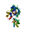
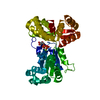
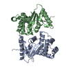
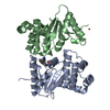
- PDB-6zui: Crystal structure of the Cys-Ser mutant of the cpYFP-based biosen... -
+ Open data

- Basic information
| Entry | Database: PDB / ID: 6zui | ||||||
|---|---|---|---|---|---|---|---|
| Title | Crystal structure of the Cys-Ser mutant of the cpYFP-based biosensor for hypochlorous acid | ||||||
 Components Components | HTH-type transcriptional repressor NemR,Green fluorescent protein,Green fluorescent protein,HTH-type transcriptional repressor NemR | ||||||
 Keywords Keywords | FLUORESCENT PROTEIN / biosensor / hypohalous acid / cpYFP / yellow fluorescent protein | ||||||
| Function / homology |  Function and homology information Function and homology informationbioluminescence / generation of precursor metabolites and energy / DNA-binding transcription factor activity / negative regulation of DNA-templated transcription / DNA binding Similarity search - Function | ||||||
| Biological species |    Aequorea victoria (jellyfish) Aequorea victoria (jellyfish) | ||||||
| Method |  X-RAY DIFFRACTION / SYNCHROTRON / MOLECULAR REPLACEMENT / Resolution: 2.20008218184 Å X-RAY DIFFRACTION / SYNCHROTRON / MOLECULAR REPLACEMENT / Resolution: 2.20008218184 Å | ||||||
 Authors Authors | Tossounian, M.A. / Van Molle, I. / Messens, J. | ||||||
| Funding support |  Belgium, 1items Belgium, 1items
| ||||||
 Citation Citation | Journal: Nat Commun / Year: 2022 Title: Hypocrates is a genetically encoded fluorescent biosensor for (pseudo)hypohalous acids and their derivatives. Authors: Kostyuk, A.I. / Tossounian, M.A. / Panova, A.S. / Thauvin, M. / Raevskii, R.I. / Ezerina, D. / Wahni, K. / Van Molle, I. / Sergeeva, A.D. / Vertommen, D. / Gorokhovatsky, A.Y. / Baranov, M.S. ...Authors: Kostyuk, A.I. / Tossounian, M.A. / Panova, A.S. / Thauvin, M. / Raevskii, R.I. / Ezerina, D. / Wahni, K. / Van Molle, I. / Sergeeva, A.D. / Vertommen, D. / Gorokhovatsky, A.Y. / Baranov, M.S. / Vriz, S. / Messens, J. / Bilan, D.S. / Belousov, V.V. #1: Journal: Biorxiv / Year: 2021Title: Monitoring oxidative inflammatory processes in live cells and tissue with Hypocrates, a genetically encoded biosensor for hypochlorite Authors: Kostyuk, A.I. / Tossounian, M.A. / Panova, A.S. / Thauvin, M. / Wahni, K. / Van Molle, I. / Raevskii, R.I. / Baranov, M.S. / Vriz, S. / Messens, J. / Bilan, D.S. / Belousov, V.V. | ||||||
| History |
|
- Structure visualization
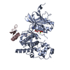
| Structure viewer | Molecule: MolmilJmol/JSmol |
|---|
- Downloads & links
-Download
| PDBx/mmCIF format | 6zui.cif.gz | 124 KB | Display | PDBx/mmCIF format |
|---|---|---|---|---|
| PDB format | pdb6zui.ent.gz | 76.2 KB | Display | PDB format |
| PDBx/mmJSON format | 6zui.json.gz | Tree view | PDBx/mmJSON format | |
| Others |  Other downloads Other downloads |
-Validation report
| Arichive directory | https://data.pdbj.org/pub/pdb/validation_reports/zu/6zuiftp://data.pdbj.org/pub/pdb/validation_reports/zu/6zui | HTTPS FTP |
|---|
-Related structure data


-Links
 PDBj
PDBj

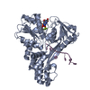
- Assembly
| Deposited unit | 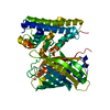
| ||||||||||||
|---|---|---|---|---|---|---|---|---|---|---|---|---|---|
| 1 |
| ||||||||||||
| Unit cell |
| ||||||||||||
| Components on special symmetry positions |
|
-Components
| #1: Protein | Mass: 49632.641 Da / Num. of mol.: 1 / Mutation: C353S Source method: isolated from a genetically manipulated source Details: The protein contains a chromophore (CR2 = Gly-Tyr-Gly) within the cpYFP domain.,The protein contains a chromophore (CR2 = Gly-Tyr-Gly) within the cpYFP domain.,The protein contains a ...Details: The protein contains a chromophore (CR2 = Gly-Tyr-Gly) within the cpYFP domain.,The protein contains a chromophore (CR2 = Gly-Tyr-Gly) within the cpYFP domain.,The protein contains a chromophore (CR2 = Gly-Tyr-Gly) within the cpYFP domain.,The protein contains a chromophore (CR2 = Gly-Tyr-Gly) within the cpYFP domain. Source: (gene. exp.) Aequorea victoria (jellyfish)Strain: K12 / Gene: nemR, ydhM, b1649, JW5874, GFP / Production host: |
|---|---|
| #2: Water | ChemComp-HOH /  Mass: 18.015 Da / Num. of mol.: 185 / Source method: isolated from a natural source / Formula: H2O Mass: 18.015 Da / Num. of mol.: 185 / Source method: isolated from a natural source / Formula: H2O |
| Has ligand of interest | Y |
| Has protein modification | Y |
-Experimental details
-Experiment
| Experiment | Method: X-RAY DIFFRACTION / Number of used crystals: 1 |
|---|
- Sample preparation
| Crystal | Density Matthews: 2.21 Å3/Da / Density % sol: 44.4 % |
|---|---|
| Crystal grow | Temperature: 283.15 K / Method: vapor diffusion, hanging drop / pH: 8 Details: Tris (0.1 M, pH 8), CaCl2 (0.1 M), MgCl2 (0.1 M) and PE15/4 (15%) Protein concentration:7 mg/mL |
-Data collection
| Diffraction | Mean temperature: 100 K / Serial crystal experiment: N |
|---|---|
| Diffraction source | Source: SYNCHROTRON / Site: SOLEIL  / Beamline: PROXIMA 2 / Wavelength: 0.980113 Å / Beamline: PROXIMA 2 / Wavelength: 0.980113 Å |
| Detector | Type: DECTRIS EIGER X 9M / Detector: PIXEL / Date: Mar 19, 2019 |
| Radiation | Protocol: SINGLE WAVELENGTH / Monochromatic (M) / Laue (L): M / Scattering type: x-ray |
| Radiation wavelength | Wavelength: 0.980113 Å / Relative weight: 1 |
| Reflection | Resolution: 2.09962866868→47.72 Å / Num. obs: 22368 / % possible obs: 83 % / Redundancy: 9.3 % / Biso Wilson estimate: 34.3012936225 Å2 / CC1/2: 0.999 / Rmerge(I) obs: 0.079 / Net I/σ(I): 16.1 |
| Reflection shell | Resolution: 2.1→2.15 Å / Rmerge(I) obs: 0.62 / Num. unique obs: 924 / CC1/2: 0.814 |
- Processing
| Software |
| |||||||||||||||||||||||||||||||||||||||||||||||||||||||||||||||
|---|---|---|---|---|---|---|---|---|---|---|---|---|---|---|---|---|---|---|---|---|---|---|---|---|---|---|---|---|---|---|---|---|---|---|---|---|---|---|---|---|---|---|---|---|---|---|---|---|---|---|---|---|---|---|---|---|---|---|---|---|---|---|---|---|
| Refinement | Method to determine structure: MOLECULAR REPLACEMENT Starting model: 4YZE, 3O77 Resolution: 2.20008218184→47.7185 Å / SU ML: 0.329732416594 / Cross valid method: FREE R-VALUE / σ(F): 1.33787706633 / Phase error: 31.970318773 Stereochemistry target values: GeoStd + Monomer Library + CDL v1.2
| |||||||||||||||||||||||||||||||||||||||||||||||||||||||||||||||
| Solvent computation | Shrinkage radii: 0.9 Å / VDW probe radii: 1.11 Å / Solvent model: FLAT BULK SOLVENT MODEL | |||||||||||||||||||||||||||||||||||||||||||||||||||||||||||||||
| Displacement parameters | Biso mean: 42.4430328993 Å2 | |||||||||||||||||||||||||||||||||||||||||||||||||||||||||||||||
| Refinement step | Cycle: LAST / Resolution: 2.20008218184→47.7185 Å
| |||||||||||||||||||||||||||||||||||||||||||||||||||||||||||||||
| Refine LS restraints |
| |||||||||||||||||||||||||||||||||||||||||||||||||||||||||||||||
| LS refinement shell |
|
