 Movie
Movie Controller

[English] 日本語
 Yorodumi
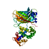
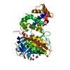
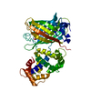
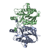
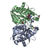
- PDB-6zsn: Crystal structure of rsGCaMP double mutant Ile80His/Val116Ile in ... -
+ Open data

- Basic information
| Entry | Database: PDB / ID: 6zsn | ||||||
|---|---|---|---|---|---|---|---|
| Title | Crystal structure of rsGCaMP double mutant Ile80His/Val116Ile in the OFF state (illuminated) | ||||||
 Components Components | Green fluorescent protein,Green fluorescent protein,Calmodulin | ||||||
 Keywords Keywords | FLUORESCENT PROTEIN / reversible switchable fluorescent protein / calcium sensor / calmodulin | ||||||
| Function / homology |  Function and homology information Function and homology informationmyosin II complex / bioluminescence / generation of precursor metabolites and energy / calcium ion binding Similarity search - Function | ||||||
| Biological species |   Aequorea victoria (jellyfish)Entacmaea quadricolor (sea anemone) Aequorea victoria (jellyfish)Entacmaea quadricolor (sea anemone) | ||||||
| Method |  X-RAY DIFFRACTION / SYNCHROTRON / MOLECULAR REPLACEMENT / Resolution: 2.6 Å X-RAY DIFFRACTION / SYNCHROTRON / MOLECULAR REPLACEMENT / Resolution: 2.6 Å | ||||||
 Authors Authors | Janowski, R. / Fuenzalida-Werner, J.P. / Mishra, K. / Stiel, A.C. / Niessing, D. | ||||||
| Funding support |  Germany, 1items Germany, 1items
| ||||||
 Citation Citation | Journal: Nat.Biotechnol. / Year: 2022 Title: Genetically encoded photo-switchable molecular sensors for optoacoustic and super-resolution imaging. Authors: Mishra, K. / Fuenzalida-Werner, J.P. / Pennacchietti, F. / Janowski, R. / Chmyrov, A. / Huang, Y. / Zakian, C. / Klemm, U. / Testa, I. / Niessing, D. / Ntziachristos, V. / Stiel, A.C. | ||||||
| History |
|
- Structure visualization
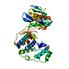
| Structure viewer | Molecule: MolmilJmol/JSmol |
|---|
- Downloads & links
-Download
| PDBx/mmCIF format | 6zsn.cif.gz | 100.8 KB | Display | PDBx/mmCIF format |
|---|---|---|---|---|
| PDB format | pdb6zsn.ent.gz | 73.7 KB | Display | PDB format |
| PDBx/mmJSON format | 6zsn.json.gz | Tree view | PDBx/mmJSON format | |
| Others |  Other downloads Other downloads |
-Validation report
| Arichive directory | https://data.pdbj.org/pub/pdb/validation_reports/zs/6zsnftp://data.pdbj.org/pub/pdb/validation_reports/zs/6zsn | HTTPS FTP |
|---|
-Related structure data
| Related structure data |  6tv7C  6ya9SC  6zsmC  7augC S: Starting model for refinement C: citing same article ( |
|---|---|
| Similar structure data |


-Links
 PDBj
PDBj


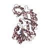
- Assembly
| Deposited unit | 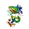
| |||||||||
|---|---|---|---|---|---|---|---|---|---|---|
| 1 |
| |||||||||
| Unit cell |
| |||||||||
| Components on special symmetry positions |
|
-Components
| #1: Protein | Mass: 47324.988 Da / Num. of mol.: 1 Source method: isolated from a genetically manipulated source Source: (gene. exp.) Aequorea victoria (jellyfish), (gene. exp.) Entacmaea quadricolor (sea anemone)Gene: GFP / Production host:  | ||||||||
|---|---|---|---|---|---|---|---|---|---|
| #2: Chemical | ChemComp-FMT /   Mass: 46.025 Da / Num. of mol.: 6 / Source method: obtained synthetically / Formula: CH2O2 Mass: 46.025 Da / Num. of mol.: 6 / Source method: obtained synthetically / Formula: CH2O2#3: Chemical | ChemComp-CA /   Mass: 40.078 Da / Num. of mol.: 4 / Source method: obtained synthetically / Formula: Ca Mass: 40.078 Da / Num. of mol.: 4 / Source method: obtained synthetically / Formula: Ca#4: Chemical | ChemComp-NA / |   Mass: 22.990 Da / Num. of mol.: 1 / Source method: obtained synthetically / Formula: Na Mass: 22.990 Da / Num. of mol.: 1 / Source method: obtained synthetically / Formula: Na#5: Water | ChemComp-HOH / |  Mass: 18.015 Da / Num. of mol.: 108 / Source method: isolated from a natural source / Formula: H2O Mass: 18.015 Da / Num. of mol.: 108 / Source method: isolated from a natural source / Formula: H2OHas ligand of interest | Y | |
-Experimental details
-Experiment
| Experiment | Method: X-RAY DIFFRACTION / Number of used crystals: 1 |
|---|
- Sample preparation
| Crystal | Density Matthews: 3.6 Å3/Da / Density % sol: 65.81 % |
|---|---|
| Crystal grow | Temperature: 292 K / Method: vapor diffusion, hanging drop / pH: 8.5 Details: 0.20 M sodium formate, 0.1 M Bis-Tris-Propane buffer pH 8.5, 19% (w/v) PEG 3350 |
-Data collection
| Diffraction | Mean temperature: 100 K / Serial crystal experiment: N |
|---|---|
| Diffraction source | Source: SYNCHROTRON / Site: SLS  / Beamline: X06DA / Wavelength: 1 Å / Beamline: X06DA / Wavelength: 1 Å |
| Detector | Type: DECTRIS PILATUS 2M-F / Detector: PIXEL / Date: Jun 25, 2020 |
| Radiation | Protocol: SINGLE WAVELENGTH / Monochromatic (M) / Laue (L): M / Scattering type: x-ray |
| Radiation wavelength | Wavelength: 1 Å / Relative weight: 1 |
| Reflection | Resolution: 2.6→50 Å / Num. obs: 21798 / % possible obs: 100 % / Redundancy: 13 % / Biso Wilson estimate: 63 Å2 / CC1/2: 0.998 / Net I/σ(I): 11.8 |
| Reflection shell | Resolution: 2.6→2.67 Å / Redundancy: 12.5 % / Mean I/σ(I) obs: 1.6 / Num. unique obs: 1571 / CC1/2: 0.616 / % possible all: 100 |
- Processing
| Software |
| ||||||||||||||||||||||||||||||||||||||||||||||||||||||||||||
|---|---|---|---|---|---|---|---|---|---|---|---|---|---|---|---|---|---|---|---|---|---|---|---|---|---|---|---|---|---|---|---|---|---|---|---|---|---|---|---|---|---|---|---|---|---|---|---|---|---|---|---|---|---|---|---|---|---|---|---|---|---|
| Refinement | Method to determine structure: MOLECULAR REPLACEMENT Starting model: 6ya9 Resolution: 2.6→46.61 Å / Cor.coef. Fo:Fc: 0.951 / Cor.coef. Fo:Fc free: 0.911 / SU B: 10.694 / SU ML: 0.214 / Cross valid method: THROUGHOUT / σ(F): 0 / ESU R: 0.336 / ESU R Free: 0.274 / Stereochemistry target values: MAXIMUM LIKELIHOOD Details: HYDROGENS HAVE BEEN ADDED IN THE RIDING POSITIONS U VALUES : REFINED INDIVIDUALLY
| ||||||||||||||||||||||||||||||||||||||||||||||||||||||||||||
| Solvent computation | Ion probe radii: 0.8 Å / Shrinkage radii: 0.8 Å / VDW probe radii: 1.2 Å / Solvent model: MASK | ||||||||||||||||||||||||||||||||||||||||||||||||||||||||||||
| Displacement parameters | Biso max: 184.86 Å2 / Biso mean: 59.2 Å2 / Biso min: 35.08 Å2
| ||||||||||||||||||||||||||||||||||||||||||||||||||||||||||||
| Refinement step | Cycle: final / Resolution: 2.6→46.61 Å
| ||||||||||||||||||||||||||||||||||||||||||||||||||||||||||||
| Refine LS restraints |
| ||||||||||||||||||||||||||||||||||||||||||||||||||||||||||||
| LS refinement shell | Resolution: 2.6→2.667 Å / Rfactor Rfree error: 0 / Total num. of bins used: 20
|
