 Movie
Movie Controller

[English] 日本語
 Yorodumi
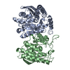
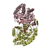
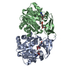
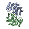
- PDB-6zl7: CRYSTAL STRUCTURE OF C173S MUTATION IN THE PMGL2 ESTERASE FROM PE... -
+ Open data

- Basic information
| Entry | Database: PDB / ID: 6zl7 | ||||||
|---|---|---|---|---|---|---|---|
| Title | CRYSTAL STRUCTURE OF C173S MUTATION IN THE PMGL2 ESTERASE FROM PERMAFROST METAGENOMIC LIBRARY | ||||||
 Components Components | PMGL2 | ||||||
 Keywords Keywords | HYDROLASE / PMGL2 / ESTERASE / PERMAFROST / METAGENOMIC LIBRARY / LIPASE / MUTANT | ||||||
| Function / homology | : / Alpha/beta hydrolase fold-3 / alpha/beta hydrolase fold / triacylglycerol lipase / triacylglycerol lipase activity / Alpha/Beta hydrolase fold / Chem-PG6 / PMGL2 Function and homology information Function and homology information | ||||||
| Biological species |  uncultured bacterium (environmental samples) uncultured bacterium (environmental samples) | ||||||
| Method |  X-RAY DIFFRACTION / SYNCHROTRON / MOLECULAR REPLACEMENT / molecular replacement / Resolution: 1.5 Å X-RAY DIFFRACTION / SYNCHROTRON / MOLECULAR REPLACEMENT / molecular replacement / Resolution: 1.5 Å | ||||||
 Authors Authors | Goryaynova, D.A. / Boyko, K.M. / Nikolaeva, A.Y. / Korzhenevskiy, D.A. / Kryukova, M.V. / Petrovskaya, L.E. / Novototskaya-Vlasova, K.A. / Rivkina, E.M. / Dolgikh, D.A. / Kirpichnikov, M.P. / Popov, V.O. | ||||||
| Funding support |  Russian Federation, 1items Russian Federation, 1items
| ||||||
 Citation Citation | Journal: To Be Published Title: CRYSTAL STRUCTURE OF C173S MUTATION IN THE PMGL2 ESTERASE FROM PERMAFROST METAGENOMIC LIBRARY Authors: Goryaynova, D.A. / Boyko, K.M. / Nikolaeva, A.Y. / Korzhenevskiy, D.A. / Kryukova, M.V. / Petrovskaya, L.E. / Novototskaya-Vlasova, K.A. / Rivkina, E.M. / Dolgikh, D.A. / Kirpichnikov, M.P. / Popov, V.O. | ||||||
| History |
|
- Structure visualization
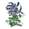
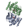
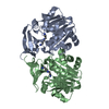
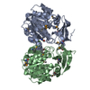
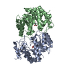
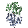
| Structure viewer | Molecule: MolmilJmol/JSmol |
|---|
- Downloads & links
-Download
| PDBx/mmCIF format | 6zl7.cif.gz | 143.1 KB | Display | PDBx/mmCIF format |
|---|---|---|---|---|
| PDB format | pdb6zl7.ent.gz | 109.2 KB | Display | PDB format |
| PDBx/mmJSON format | 6zl7.json.gz | Tree view | PDBx/mmJSON format | |
| Others |  Other downloads Other downloads |
-Validation report
| Arichive directory | https://data.pdbj.org/pub/pdb/validation_reports/zl/6zl7ftp://data.pdbj.org/pub/pdb/validation_reports/zl/6zl7 | HTTPS FTP |
|---|
-Related structure data
| Related structure data |  6qinS S: Starting model for refinement |
|---|---|
| Similar structure data |
-Links
 PDBj
PDBj
- Assembly
| Deposited unit | 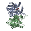
| ||||||||
|---|---|---|---|---|---|---|---|---|---|
| 1 |
| ||||||||
| Unit cell |
|
-Components
| #1: Protein | Mass: 37546.379 Da / Num. of mol.: 2 / Mutation: T173S Source method: isolated from a genetically manipulated source Source: (gene. exp.) uncultured bacterium (environmental samples)Production host: #2: Chemical | ChemComp-MG / |   Mass: 24.305 Da / Num. of mol.: 1 / Source method: obtained synthetically / Formula: Mg Mass: 24.305 Da / Num. of mol.: 1 / Source method: obtained synthetically / Formula: Mg#3: Chemical | 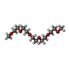  Mass: 266.331 Da / Num. of mol.: 2 / Source method: obtained synthetically / Formula: C12H26O6 Mass: 266.331 Da / Num. of mol.: 2 / Source method: obtained synthetically / Formula: C12H26O6#4: Water | ChemComp-HOH / |  Mass: 18.015 Da / Num. of mol.: 453 / Source method: isolated from a natural source / Formula: H2O Mass: 18.015 Da / Num. of mol.: 453 / Source method: isolated from a natural source / Formula: H2OHas ligand of interest | N | |
|---|
-Experimental details
-Experiment
| Experiment | Method: X-RAY DIFFRACTION / Number of used crystals: 1 |
|---|
- Sample preparation
| Crystal | Density Matthews: 2.07 Å3/Da / Density % sol: 40.62 % |
|---|---|
| Crystal grow | Temperature: 288 K / Method: vapor diffusion, hanging drop / pH: 7.5 Details: 250mM Magnesium chloride, 12-18% PEG3350, 100mM HEPES pH 7.5 |
-Data collection
| Diffraction | Mean temperature: 100 K / Serial crystal experiment: N | ||||||||||||||||||||||||||||||
|---|---|---|---|---|---|---|---|---|---|---|---|---|---|---|---|---|---|---|---|---|---|---|---|---|---|---|---|---|---|---|---|
| Diffraction source | Source: SYNCHROTRON / Site: SPring-8  / Beamline: BL41XU / Wavelength: 1 Å / Beamline: BL41XU / Wavelength: 1 Å | ||||||||||||||||||||||||||||||
| Detector | Type: DECTRIS EIGER X 16M / Detector: PIXEL / Date: Jun 29, 2017 | ||||||||||||||||||||||||||||||
| Radiation | Protocol: SINGLE WAVELENGTH / Monochromatic (M) / Laue (L): M / Scattering type: x-ray | ||||||||||||||||||||||||||||||
| Radiation wavelength | Wavelength: 1 Å / Relative weight: 1 | ||||||||||||||||||||||||||||||
| Reflection | Resolution: 1.5→56.54 Å / Num. obs: 95216 / % possible obs: 97.6 % / Redundancy: 6.3 % / CC1/2: 0.999 / Rmerge(I) obs: 0.067 / Rpim(I) all: 0.029 / Rrim(I) all: 0.073 / Net I/σ(I): 16.1 | ||||||||||||||||||||||||||||||
| Reflection shell | Diffraction-ID: 1
|
-Phasing
| Phasing | Method: molecular replacement |
|---|
- Processing
| Software |
| |||||||||||||||||||||||||||||||||||||||||||||
|---|---|---|---|---|---|---|---|---|---|---|---|---|---|---|---|---|---|---|---|---|---|---|---|---|---|---|---|---|---|---|---|---|---|---|---|---|---|---|---|---|---|---|---|---|---|---|
| Refinement | Method to determine structure: MOLECULAR REPLACEMENT Starting model: 6QIN Resolution: 1.5→56.54 Å / Cor.coef. Fo:Fc: 0.966 / Cor.coef. Fo:Fc free: 0.956 / SU B: 1.302 / SU ML: 0.048 / SU R Cruickshank DPI: 0.0718 / Cross valid method: THROUGHOUT / σ(F): 0 / ESU R: 0.072 / ESU R Free: 0.073 / Stereochemistry target values: MAXIMUM LIKELIHOOD / Details: U VALUES : REFINED INDIVIDUALLY
| |||||||||||||||||||||||||||||||||||||||||||||
| Solvent computation | Ion probe radii: 0.8 Å / Shrinkage radii: 0.8 Å / VDW probe radii: 1.2 Å / Solvent model: MASK | |||||||||||||||||||||||||||||||||||||||||||||
| Displacement parameters | Biso max: 89.9 Å2 / Biso mean: 18.289 Å2 / Biso min: 7.38 Å2
| |||||||||||||||||||||||||||||||||||||||||||||
| Refinement step | Cycle: final / Resolution: 1.5→56.54 Å
| |||||||||||||||||||||||||||||||||||||||||||||
| Refine LS restraints |
| |||||||||||||||||||||||||||||||||||||||||||||
| LS refinement shell | Resolution: 1.5→1.539 Å / Rfactor Rfree error: 0 / Total num. of bins used: 20
|
