 Movie
Movie Controller

[English] 日本語
 Yorodumi
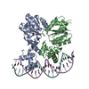
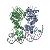
- PDB-6zj2: Structure of RcsB from Salmonella enterica serovar Typhimurium bo... -
+ Open data

- Basic information
| Entry | Database: PDB / ID: 6zj2 | |||||||||||||||
|---|---|---|---|---|---|---|---|---|---|---|---|---|---|---|---|---|
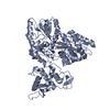
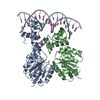
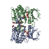
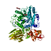
| Title | Structure of RcsB from Salmonella enterica serovar Typhimurium bound to promoter rprA in the presence of phosphomimetic BeF3- | |||||||||||||||
 Components Components |
| |||||||||||||||
 Keywords Keywords | DNA BINDING PROTEIN / response regulator / phosphorylation / two-component systems / transcriptional factor | |||||||||||||||
| Function / homology |  Function and homology information Function and homology informationphosphorelay signal transduction system / transcription cis-regulatory region binding / DNA-binding transcription factor activity / regulation of DNA-templated transcription Similarity search - Function | |||||||||||||||
| Biological species |  Salmonella enterica subsp. enterica serovar Typhimurium str. LT2 (bacteria)Salmonella enterica subsp. enterica serovar Typhimurium (bacteria) Salmonella enterica subsp. enterica serovar Typhimurium str. LT2 (bacteria)Salmonella enterica subsp. enterica serovar Typhimurium (bacteria) | |||||||||||||||
| Method |  X-RAY DIFFRACTION / SYNCHROTRON / MOLECULAR REPLACEMENT / Resolution: 3.38 Å X-RAY DIFFRACTION / SYNCHROTRON / MOLECULAR REPLACEMENT / Resolution: 3.38 Å | |||||||||||||||
 Authors Authors | Huesa, J. / Marina, A. / Casino, P. | |||||||||||||||
| Funding support |  Spain, 4items Spain, 4items
| |||||||||||||||
 Citation Citation | Journal: Nucleic Acids Res. / Year: 2021 Title: Structure-based analyses of Salmonella RcsB variants unravel new features of the Rcs regulon. Authors: Huesa, J. / Giner-Lamia, J. / Pucciarelli, M.G. / Paredes-Martinez, F. / Portillo, F.G. / Marina, A. / Casino, P. | |||||||||||||||
| History |
|
- Structure visualization
| Structure viewer | Molecule: MolmilJmol/JSmol |
|---|
- Downloads & links
-Download
| PDBx/mmCIF format | 6zj2.cif.gz | 320.1 KB | Display | PDBx/mmCIF format |
|---|---|---|---|---|
| PDB format | pdb6zj2.ent.gz | 247.8 KB | Display | PDB format |
| PDBx/mmJSON format | 6zj2.json.gz | Tree view | PDBx/mmJSON format | |
| Others |  Other downloads Other downloads |
-Validation report
| Arichive directory | https://data.pdbj.org/pub/pdb/validation_reports/zj/6zj2ftp://data.pdbj.org/pub/pdb/validation_reports/zj/6zj2 | HTTPS FTP |
|---|
-Related structure data
| Related structure data | 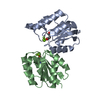 6ziiC 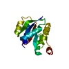 6zilC  6zixC 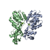 5o8zS C: citing same article ( S: Starting model for refinement |
|---|---|
| Similar structure data |


-Links
 PDBj
PDBj


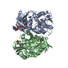
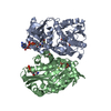
- Assembly
| Deposited unit | 
| ||||||||
|---|---|---|---|---|---|---|---|---|---|
| 1 | 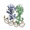
| ||||||||
| 2 | 
| ||||||||
| 3 | 
| ||||||||
| Unit cell |
|
-Components
| #1: Protein | Mass: 23743.650 Da / Num. of mol.: 8 Source method: isolated from a genetically manipulated source Source: (gene. exp.) Salmonella enterica subsp. enterica serovar Typhimurium str. LT2 (bacteria)Gene: rcsB, STM2270 / Production host: #2: DNA chain | Mass: 7080.584 Da / Num. of mol.: 2 Source method: isolated from a genetically manipulated source Source: (gene. exp.) Salmonella enterica subsp. enterica serovar Typhimurium (bacteria)Production host: #3: DNA chain | Mass: 7040.561 Da / Num. of mol.: 2 Source method: isolated from a genetically manipulated source Source: (gene. exp.) Salmonella enterica subsp. enterica serovar Typhimurium (bacteria)Production host: #4: Chemical |   Mass: 66.007 Da / Num. of mol.: 2 / Source method: obtained synthetically / Formula: BeF3 Mass: 66.007 Da / Num. of mol.: 2 / Source method: obtained synthetically / Formula: BeF3#5: Chemical | ChemComp-MG /   Mass: 24.305 Da / Num. of mol.: 4 / Source method: obtained synthetically / Formula: Mg Mass: 24.305 Da / Num. of mol.: 4 / Source method: obtained synthetically / Formula: Mg |
|---|
-Experimental details
-Experiment
| Experiment | Method: X-RAY DIFFRACTION / Number of used crystals: 1 |
|---|
- Sample preparation
| Crystal | Density Matthews: 2.59 Å3/Da / Density % sol: 52.55 % |
|---|---|
| Crystal grow | Temperature: 294 K / Method: vapor diffusion, sitting drop / Details: 15% PEG 8000, MES pH 6.5 and 0.2M NaAc |
-Data collection
| Diffraction | Mean temperature: 100 K / Serial crystal experiment: N | |||||||||||||||||||||||||||
|---|---|---|---|---|---|---|---|---|---|---|---|---|---|---|---|---|---|---|---|---|---|---|---|---|---|---|---|---|
| Diffraction source | Source: SYNCHROTRON / Site: ALBA / Beamline: XALOC / Wavelength: 0.97949 Å | |||||||||||||||||||||||||||
| Detector | Type: DECTRIS PILATUS3 6M / Detector: PIXEL / Date: Sep 26, 2019 | |||||||||||||||||||||||||||
| Radiation | Protocol: SINGLE WAVELENGTH / Monochromatic (M) / Laue (L): M / Scattering type: x-ray | |||||||||||||||||||||||||||
| Radiation wavelength | Wavelength: 0.97949 Å / Relative weight: 1 | |||||||||||||||||||||||||||
| Reflection | Resolution: 3.38→45.99 Å / Num. obs: 30572 / % possible obs: 99.1 % / Redundancy: 3.8 % / CC1/2: 0.983 / Rmerge(I) obs: 0.228 / Rpim(I) all: 0.133 / Rrim(I) all: 0.266 / Net I/σ(I): 4.2 | |||||||||||||||||||||||||||
| Reflection shell | Diffraction-ID: 1 / Redundancy: 3.6 %
|
- Processing
| Software |
| ||||||||||||||||||||||||||||||||||||||||||||||||||||||||||||
|---|---|---|---|---|---|---|---|---|---|---|---|---|---|---|---|---|---|---|---|---|---|---|---|---|---|---|---|---|---|---|---|---|---|---|---|---|---|---|---|---|---|---|---|---|---|---|---|---|---|---|---|---|---|---|---|---|---|---|---|---|---|
| Refinement | Method to determine structure: MOLECULAR REPLACEMENT Starting model: 5O8Z Resolution: 3.38→45.99 Å / Cor.coef. Fo:Fc: 0.866 / Cor.coef. Fo:Fc free: 0.829 / SU B: 22.271 / SU ML: 0.389 / Cross valid method: THROUGHOUT / σ(F): 0 / ESU R Free: 0.151 / Stereochemistry target values: MAXIMUM LIKELIHOOD Details: HYDROGENS HAVE BEEN ADDED IN THE RIDING POSITIONS U VALUES : REFINED INDIVIDUALLY
| ||||||||||||||||||||||||||||||||||||||||||||||||||||||||||||
| Solvent computation | Ion probe radii: 0.8 Å / Shrinkage radii: 0.8 Å / VDW probe radii: 1.2 Å / Solvent model: MASK | ||||||||||||||||||||||||||||||||||||||||||||||||||||||||||||
| Displacement parameters | Biso max: 195.64 Å2 / Biso mean: 85.244 Å2 / Biso min: 35.82 Å2
| ||||||||||||||||||||||||||||||||||||||||||||||||||||||||||||
| Refinement step | Cycle: final / Resolution: 3.38→45.99 Å
| ||||||||||||||||||||||||||||||||||||||||||||||||||||||||||||
| Refine LS restraints |
| ||||||||||||||||||||||||||||||||||||||||||||||||||||||||||||
| LS refinement shell | Resolution: 3.38→3.468 Å / Rfactor Rfree error: 0 / Total num. of bins used: 20
|
