 Movie
Movie Controller

[English] 日本語
 Yorodumi
- PDB-6zbg: Merozoite surface protein 1 (MSP-1) from Plasmodium falciparum, a... -
+ Open data

- Basic information
| Entry | Database: PDB / ID: 6zbg | ||||||
|---|---|---|---|---|---|---|---|
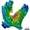
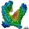
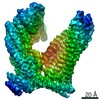
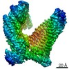
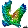
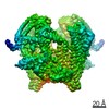
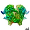
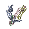
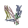
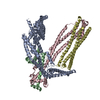
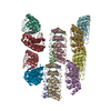
| Title | Merozoite surface protein 1 (MSP-1) from Plasmodium falciparum, alternative conformation 4 | ||||||
 Components Components |
| ||||||
 Keywords Keywords | MEMBRANE PROTEIN / Merozoite surface protein 1 / malaria / Plasmodium falciparum / MSP-1 / p190 / GPI-anchored membrane protein | ||||||
| Function / homology |  Function and homology information Function and homology informationvacuolar membrane / side of membrane / extracellular region / plasma membrane Similarity search - Function | ||||||
| Biological species |  | ||||||
| Method | ELECTRON MICROSCOPY / single particle reconstruction / cryo EM / Resolution: 3.2 Å | ||||||
 Authors Authors | Dijkman, P.M. / Kudryashev, M. | ||||||
| Funding support |  Germany, 1items Germany, 1items
| ||||||
 Citation Citation | Journal: Sci Adv / Year: 2021 Title: Structure of the merozoite surface protein 1 from . Authors: Patricia M Dijkman / Tanja Marzluf / Yingyi Zhang / Shih-Ying Scott Chang / Dominic Helm / Michael Lanzer / Hermann Bujard / Mikhail Kudryashev / Abstract: The merozoite surface protein 1 (MSP-1) is the most abundant protein on the surface of the erythrocyte-invading merozoite, the causative agent of malaria. MSP-1 is essential for merozoite formation, ...The merozoite surface protein 1 (MSP-1) is the most abundant protein on the surface of the erythrocyte-invading merozoite, the causative agent of malaria. MSP-1 is essential for merozoite formation, entry into and escape from erythrocytes, and is a promising vaccine candidate. Here, we present monomeric and dimeric structures of full-length MSP-1. MSP-1 adopts an unusual fold with a large central cavity. Its fold includes several coiled-coils and shows structural homology to proteins associated with membrane and cytoskeleton interactions. MSP-1 formed dimers through these domains in a concentration-dependent manner. Dimerization is affected by the presence of the erythrocyte cytoskeleton protein spectrin, which may compete for the dimerization interface. Our work provides structural insights into the possible mode of interaction of MSP-1 with erythrocytes and establishes a framework for future investigations into the role of MSP-1 in infection and immunity. #1: Journal: Sci Adv / Year: 2021Title: Structure of the merozoite surface protein 1 from Plasmodium falciparum Authors: Dijkman, P.M. / Marzluf, T. / Zhang, Y. / Chang, S.Y.S. / Helm, D. / Lanzer, M. / Bujard, H. / Kudryashev, M. | ||||||
| History |
|
- Structure visualization
| Movie |
Movie viewer |
|---|---|
| Structure viewer | Molecule: MolmilJmol/JSmol |
- Downloads & links
-Download
| PDBx/mmCIF format | 6zbg.cif.gz | 419.2 KB | Display | PDBx/mmCIF format |
|---|---|---|---|---|
| PDB format | pdb6zbg.ent.gz | 338.3 KB | Display | PDB format |
| PDBx/mmJSON format | 6zbg.json.gz | Tree view | PDBx/mmJSON format | |
| Others |  Other downloads Other downloads |
-Validation report
| Summary document | 6zbg_validation.pdf.gz | 1.4 MB | Display | wwPDB validaton report |
|---|---|---|---|---|
| Full document | 6zbg_full_validation.pdf.gz | 1.4 MB | Display | |
| Data in XML | 6zbg_validation.xml.gz | 43.7 KB | Display | |
| Data in CIF | 6zbg_validation.cif.gz | 65.6 KB | Display | |
| Arichive directory | https://data.pdbj.org/pub/pdb/validation_reports/zb/6zbgftp://data.pdbj.org/pub/pdb/validation_reports/zb/6zbg | HTTPS FTP |
-Related structure data
| Related structure data | 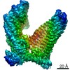 11154MC 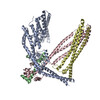 6zbcC 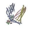 6zbdC 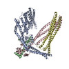 6zbeC 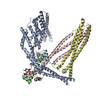 6zbfC 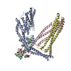 6zbhC 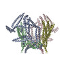 6zbjC 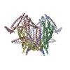 6zblC M: map data used to model this data C: citing same article ( |
|---|---|
| Similar structure data | |
| EM raw data | EMPIAR-10437 (Title: Single-particle cryo-EM of the full-length merozoite surface protein 1 from Plasmodium falciparum Data size: 34.0 TB Data #1: Unaligned movies of MSP-1 [micrographs - multiframe] Data #2: Unaligned movies of hdMSP-1 (dataset 1) [micrographs - multiframe] Data #3: Unaligned movies of hdMSP-1 (dataset 2) [micrographs - multiframe] Data #4: Unaligned movies of hdMSP-1 (dataset 3) [micrographs - multiframe] Data #5: Unaligned movies of hdMSP-1 (dataset 4) [micrographs - multiframe] Data #6: Final particle stack for hdMSP-1 conformation 1 [picked particles - single frame - processed] Data #7: Final particle stack for hdMSP-1 conformation 2 [picked particles - single frame - processed] Data #8: Final particle stack for MSP-1 main conformation [picked particles - single frame - processed] Data #9: Final particle stack for MSP-1 alternative conformation 1 [picked particles - single frame - processed] Data #10: Final particle stack for MSP-1 alternative conformation 2 [picked particles - single frame - processed] Data #11: Final particle stack for MSP-1 alternative conformation 3 [picked particles - single frame - processed] Data #12: Final particle stack for MSP-1 alternative conformation 4 [picked particles - single frame - processed] Data #13: Final particle stack for MSP-1 alternative conformation 5 [picked particles - single frame - processed]) |

-Links
 PDBj
PDBj- Assembly
| Deposited unit | 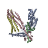
|
|---|---|
| 1 |
|
-Components
| #1: Protein | Mass: 81773.539 Da / Num. of mol.: 1 Source method: isolated from a genetically manipulated source Details: Subunit of the MSP-1 complex Source: (gene. exp.) Gene: gp190/ MSA1/ MSP1/ PMMSA / Production host:  |
|---|---|
| #2: Protein | Mass: 20217.637 Da / Num. of mol.: 1 Source method: isolated from a genetically manipulated source Details: Subunit of the MSP-1 complex Source: (gene. exp.) Gene: msp1 / Production host: |
| #3: Protein | Mass: 46390.676 Da / Num. of mol.: 1 Source method: isolated from a genetically manipulated source Details: Subunit of the MSP-1 complex Source: (gene. exp.) Gene: msp1 / Production host: |
| #4: Protein | Mass: 43243.133 Da / Num. of mol.: 1 Source method: isolated from a genetically manipulated source Details: Subunit of the MSP-1 complex Source: (gene. exp.) Gene: msp1 / Production host: |
| Has protein modification | Y |
-Experimental details
-Experiment
| Experiment | Method: ELECTRON MICROSCOPY |
|---|---|
| EM experiment | Aggregation state: PARTICLE / 3D reconstruction method: single particle reconstruction |
- Sample preparation
| Component | Name: Merozoite surface protein 1 (MSP-1) / Type: COMPLEX Details: Heteromeric assembly of p83/30 fusion and p38/42 fusion of MSP-1 from Plasmodium falciparum (monomer), processed with SUB-1 protease into the MSP-1 complex composed of the subunits p83, p30, p38, and p42 Entity ID: all / Source: RECOMBINANT |
|---|---|
| Molecular weight | Experimental value: NO |
| Source (natural) | Organism: |
| Source (recombinant) | Organism: |
| Buffer solution | pH: 7.4 |
| Specimen | Embedding applied: NO / Shadowing applied: NO / Staining applied: NO / Vitrification applied: YES Details: Heteromeric assembly of p83/30 fusion and p38/42 fusion of MSP-1 from Plasmodium falciparum (monomer), processed with SUB-1 protease into the MSP-1 complex composed of the subunits p83, p30, p38, and p42 |
| Vitrification | Cryogen name: ETHANE |
- Electron microscopy imaging
| Experimental equipment |  Model: Titan Krios / Image courtesy: FEI Company |
|---|---|
| Microscopy | Model: FEI TITAN KRIOS |
| Electron gun | Electron source:  FIELD EMISSION GUN / Accelerating voltage: 300 kV / Illumination mode: FLOOD BEAM FIELD EMISSION GUN / Accelerating voltage: 300 kV / Illumination mode: FLOOD BEAM |
| Electron lens | Mode: BRIGHT FIELD / Nominal magnification: 130000 X / Nominal defocus max: 3000 nm / Nominal defocus min: 500 nm |
| Specimen holder | Cryogen: NITROGEN / Specimen holder model: FEI TITAN KRIOS AUTOGRID HOLDER |
| Image recording | Electron dose: 64 e/Å2 / Film or detector model: GATAN K2 SUMMIT (4k x 4k) |
| Image scans | Movie frames/image: 40 |
- Processing
| EM software | Name: cryoSPARC / Version: 2.5 / Category: 3D reconstruction |
|---|---|
| CTF correction | Type: PHASE FLIPPING AND AMPLITUDE CORRECTION |
| Symmetry | Point symmetry: C1 (asymmetric) |
| 3D reconstruction | Resolution: 3.2 Å / Resolution method: FSC 0.143 CUT-OFF / Num. of particles: 116037 / Symmetry type: POINT |
