[English] 日本語
Yorodumi
- PDB-6z8v: X-ray structure of the complex between human alpha thrombin and a... -

+
Open data


ID or keywords:

Loading...

-
Basic information

Entry
Database: PDB / ID: 6z8v
Title
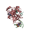
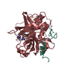
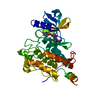
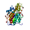
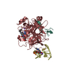
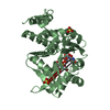
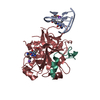
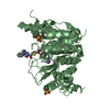
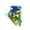
X-ray structure of the complex between human alpha thrombin and a thrombin binding aptamer variant (TBA-3L), which contains 1-beta-D-lactopyranosyl residue in the side chain of Thy3 at N3.
Components
  • (Prothrombin) x 2
  • TBA-3L
KeywordsHYDROLASE / Modified aptamer / DNA-protein complex / Anticoaugulant / G-quadruplex
Function / homology
Function and homology information


positive regulation of lipid kinase activity / positive regulation of phospholipase C-activating G protein-coupled receptor signaling pathway / cytolysis by host of symbiont cells / thrombospondin receptor activity / Defective factor XII causes hereditary angioedema / thrombin / regulation of blood coagulation / neutrophil-mediated killing of gram-negative bacterium / ligand-gated ion channel signaling pathway / Defective F8 cleavage by thrombin ...positive regulation of lipid kinase activity / positive regulation of phospholipase C-activating G protein-coupled receptor signaling pathway / cytolysis by host of symbiont cells / thrombospondin receptor activity / Defective factor XII causes hereditary angioedema / thrombin / regulation of blood coagulation / neutrophil-mediated killing of gram-negative bacterium / ligand-gated ion channel signaling pathway / Defective F8 cleavage by thrombin / Platelet Aggregation (Plug Formation) / negative regulation of astrocyte differentiation / negative regulation of platelet activation / positive regulation of collagen biosynthetic process / negative regulation of cytokine production involved in inflammatory response / positive regulation of blood coagulation / negative regulation of fibrinolysis / Gamma-carboxylation of protein precursors / Transport of gamma-carboxylated protein precursors from the endoplasmic reticulum to the Golgi apparatus / Common Pathway of Fibrin Clot Formation / Removal of aminoterminal propeptides from gamma-carboxylated proteins / regulation of cytosolic calcium ion concentration / fibrinolysis / Intrinsic Pathway of Fibrin Clot Formation / Peptide ligand-binding receptors / positive regulation of release of sequestered calcium ion into cytosol / acute-phase response / Regulation of Complement cascade / negative regulation of proteolysis / Cell surface interactions at the vascular wall / lipopolysaccharide binding / positive regulation of receptor signaling pathway via JAK-STAT / growth factor activity / positive regulation of insulin secretion / platelet activation / response to wounding / positive regulation of protein localization to nucleus / Golgi lumen / Regulation of Insulin-like Growth Factor (IGF) transport and uptake by Insulin-like Growth Factor Binding Proteins (IGFBPs) / positive regulation of reactive oxygen species metabolic process / antimicrobial humoral immune response mediated by antimicrobial peptide / blood coagulation / Thrombin signalling through proteinase activated receptors (PARs) / heparin binding / positive regulation of cell growth / regulation of cell shape / G alpha (q) signalling events / collagen-containing extracellular matrix / blood microparticle / positive regulation of phosphatidylinositol 3-kinase/protein kinase B signal transduction / cell surface receptor signaling pathway / positive regulation of protein phosphorylation / G protein-coupled receptor signaling pathway / endoplasmic reticulum lumen / serine-type endopeptidase activity / signaling receptor binding / positive regulation of cell population proliferation / calcium ion binding / proteolysis / extracellular space / extracellular exosome / extracellular region / plasma membrane
Similarity search - Function
Prothrombin/thrombin / Thrombin light chain / Thrombin light chain domain superfamily / : / Thrombin light chain / Kringle domain / Kringle / Kringle, conserved site / Kringle superfamily / Kringle domain signature. ...Prothrombin/thrombin / Thrombin light chain / Thrombin light chain domain superfamily / : / Thrombin light chain / Kringle domain / Kringle / Kringle, conserved site / Kringle superfamily / Kringle domain signature. / Kringle domain profile. / Kringle domain / Vitamin K-dependent carboxylation/gamma-carboxyglutamic (GLA) domain / Gamma-carboxyglutamic acid-rich (GLA) domain / Gamma-carboxyglutamic acid-rich (GLA) domain superfamily / Vitamin K-dependent carboxylation domain. / Gla domain profile. / Domain containing Gla (gamma-carboxyglutamate) residues. / Kringle-like fold / Serine proteases, trypsin family, histidine active site / Serine proteases, trypsin family, serine active site / Peptidase S1A, chymotrypsin family / Serine proteases, trypsin family, histidine active site. / Serine proteases, trypsin domain profile. / Serine proteases, trypsin family, serine active site. / Trypsin-like serine protease / Serine proteases, trypsin domain / Trypsin / Peptidase S1, PA clan, chymotrypsin-like fold / Peptidase S1, PA clan
Similarity search - Domain/homology
Chem-0G6 / : / DNA / DNA (> 10) / Prothrombin
Similarity search - Component
Biological speciesHomo sapiens (human)
synthetic construct (others)
MethodX-RAY DIFFRACTION / SYNCHROTRON / MOLECULAR REPLACEMENT / Resolution: 1.58 Å
AuthorsTroisi, R. / Timofeev, E.N. / Sica, F.
Funding support Russian Federation, 1items
OrganizationGrant numberCountry
Russian Foundation for Basic Research18-04-00614 Russian Federation
CitationJournal: Mol Ther Nucleic Acids / Year: 2021
Title: Expanding the recognition interface of the thrombin-binding aptamer HD1 through modification of residues T3 and T12.
Authors: Smirnov, I. / Kolganova, N. / Troisi, R. / Sica, F. / Timofeev, E.
History
DepositionJun 2, 2020Deposition site: PDBE / Processing site: PDBE
Revision 1.0Jan 27, 2021Provider: repository / Type: Initial release
Revision 1.1Mar 3, 2021Group: Database references / Category: citation
Item: _citation.country / _citation.journal_abbrev ..._citation.country / _citation.journal_abbrev / _citation.journal_id_ISSN / _citation.journal_volume / _citation.page_first / _citation.page_last / _citation.pdbx_database_id_PubMed / _citation.title
Revision 1.2Jan 24, 2024Group: Data collection / Database references / Refinement description
Category: chem_comp_atom / chem_comp_bond ...chem_comp_atom / chem_comp_bond / citation / database_2 / pdbx_initial_refinement_model
Item: _citation.country / _database_2.pdbx_DOI / _database_2.pdbx_database_accession

-
Structure visualization

Structure viewerMolecule:
MolmilJmol/JSmol

Downloads & links

-
Assembly

Deposited unit
L: Prothrombin
H: Prothrombin
A: TBA-3L
hetero molecules


Theoretical massNumber of molelcules
Total (without water)39,2176
Polymers38,7013
Non-polymers5163
Water3,639202
1


  • Idetical with deposited unit
  • defined by author&software
TypeNameSymmetry operationNumber
identity operation1_555x,y,z1
Buried area3600 Å2
ΔGint-28 kcal/mol
Surface area14030 Å2
MethodPISA
Unit cell
Length a, b, c (Å)94.916, 94.916, 125.397
Angle α, β, γ (deg.)90.000, 90.000, 120.000
Int Tables number154
Space group name H-MP3221

-
Components

-
Protein/peptide / Protein / DNA chain , 3 types, 3 molecules LHA

#1: Protein/peptide Prothrombin / Coagulation factor II


Mass: 4096.534 Da / Num. of mol.: 1 / Source method: isolated from a natural source / Source: (natural) Homo sapiens (human) / References: UniProt: P00734, thrombin
#2: Protein Prothrombin / Coagulation factor II


Mass: 29780.219 Da / Num. of mol.: 1 / Source method: isolated from a natural source / Source: (natural) Homo sapiens (human) / References: UniProt: P00734, thrombin
#3: DNA chain TBA-3L


Mass: 4824.127 Da / Num. of mol.: 1 / Source method: obtained synthetically / Source: (synth.) synthetic construct (others)

-
Non-polymers , 4 types, 205 molecules

#4: Chemical ChemComp-0G6 / D-phenylalanyl-N-[(2S,3S)-6-{[amino(iminio)methyl]amino}-1-chloro-2-hydroxyhexan-3-yl]-L-prolinamide / PPACK


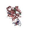
Type: peptide-like / Mass: 453.986 Da / Num. of mol.: 1 / Source method: obtained synthetically / Formula: C21H34ClN6O3
#5: Chemical ChemComp-NA / SODIUM ION


Mass: 22.990 Da / Num. of mol.: 1 / Source method: obtained synthetically / Formula: Na
#6: Chemical ChemComp-K / POTASSIUM ION


Mass: 39.098 Da / Num. of mol.: 1 / Source method: obtained synthetically / Formula: K
#7: Water ChemComp-HOH / water


Mass: 18.015 Da / Num. of mol.: 202 / Source method: isolated from a natural source / Formula: H2O

-
Details

Has ligand of interestY

-
Experimental details

-
Experiment

ExperimentMethod: X-RAY DIFFRACTION / Number of used crystals: 1

-
Sample preparation

CrystalDensity Matthews: 3.9 Å3/Da / Density % sol: 68.5 %
Crystal growTemperature: 293 K / Method: vapor diffusion, hanging drop / pH: 7.5
Details: 200 mM KCl, 35% v/v pentaerythritol propoxylate, 50 mM HEPES, pH 7.5

-
Data collection

DiffractionMean temperature: 100 K / Serial crystal experiment: N
Diffraction sourceSource: SYNCHROTRON / Site: Diamond / Beamline: I04 / Wavelength: 0.9795 Å
DetectorType: DECTRIS EIGER2 XE 16M / Detector: PIXEL / Date: Dec 2, 2019
RadiationProtocol: SINGLE WAVELENGTH / Monochromatic (M) / Laue (L): M / Scattering type: x-ray
Radiation wavelengthWavelength: 0.9795 Å / Relative weight: 1
ReflectionResolution: 1.58→82.2 Å / Num. obs: 54291 / % possible obs: 95.9 % / Redundancy: 19.6 % / CC1/2: 1 / Net I/σ(I): 23.2
Reflection shellResolution: 1.58→1.76 Å / Redundancy: 10.6 % / Mean I/σ(I) obs: 2 / Num. unique obs: 2718 / CC1/2: 0.8 / % possible all: 81.2

-
Processing

Software
NameVersionClassification
autoPROCdata processing
STARANISOdata processing
PHASERphasing
REFMAC5.8.0258refinement
PDB_EXTRACT3.25data extraction
XDSdata reduction
Aimlessdata scaling
RefinementMethod to determine structure: MOLECULAR REPLACEMENT
Starting model: 1PPB
Resolution: 1.58→82.2 Å / Cor.coef. Fo:Fc: 0.961 / Cor.coef. Fo:Fc free: 0.95 / SU B: 3.677 / SU ML: 0.055 / Cross valid method: THROUGHOUT / σ(F): 0 / ESU R: 0.09 / ESU R Free: 0.091 / Stereochemistry target values: MAXIMUM LIKELIHOOD / Details: U VALUES : WITH TLS ADDED
RfactorNum. reflection% reflectionSelection details
Rfree0.2057 2675 4.9 %RANDOM
Rwork0.1797 ---
obs0.181 51616 60.5 %-
Solvent computationIon probe radii: 0.8 Å / Shrinkage radii: 0.8 Å / VDW probe radii: 1.2 Å / Solvent model: MASK
Displacement parametersBiso max: 74.07 Å2 / Biso mean: 27.472 Å2 / Biso min: 13.15 Å2
Baniso -1Baniso -2Baniso -3
1-0 Å20 Å20 Å2
2--0 Å2-0 Å2
3----0 Å2
Refinement stepCycle: final / Resolution: 1.58→82.2 Å
ProteinNucleic acidLigandSolventTotal
Num. atoms2239 312 32 202 2785
Biso mean--19.05 33.52 -
Num. residues----294
Refine LS restraints
Refine-IDTypeDev idealDev ideal targetNumber
X-RAY DIFFRACTIONr_bond_refined_d0.0150.0122728
X-RAY DIFFRACTIONr_angle_refined_deg2.221.6053759
X-RAY DIFFRACTIONr_dihedral_angle_1_deg7.3035284
X-RAY DIFFRACTIONr_dihedral_angle_2_deg30.43621.406128
X-RAY DIFFRACTIONr_dihedral_angle_3_deg14.74915409
X-RAY DIFFRACTIONr_dihedral_angle_4_deg13.5781519
X-RAY DIFFRACTIONr_chiral_restr0.1530.2340
X-RAY DIFFRACTIONr_gen_planes_refined0.0140.021999
LS refinement shellResolution: 1.581→1.622 Å / Rfactor Rfree error: 0 / Total num. of bins used: 20
RfactorNum. reflection% reflection
Rfree0.287 5 -
Rwork0.316 166 -
all-171 -
obs--2.61 %
Refinement TLS params.

Method: refined / Refine-ID: X-RAY DIFFRACTION

IDL112)L122)L132)L222)L232)L332)S11 (Å °)S12 (Å °)S13 (Å °)S21 (Å °)S22 (Å °)S23 (Å °)S31 (Å °)S32 (Å °)S33 (Å °)T112)T122)T132)T222)T232)T332)Origin x (Å)Origin y (Å)Origin z (Å)
12.80130.02351.7851.96720.46763.2768-0.0616-0.0540.21970.0093-0.04410.2939-0.3193-0.32320.10570.0660.0160.02660.0928-0.01680.0765-60.146335.27127.6925
21.09480.05850.12571.64080.51451.68440.0382-0.011-0.08780.02220.0137-0.06560.1039-0.0044-0.05190.0144-0.01-0.00360.02080.00020.0105-48.490521.88240.0852
34.4941-0.5707-0.46632.25610.46042.02630.02420.13070.21110.0819-0.053-0.0611-0.10360.00210.02880.0803-0.05270.00570.07160.0010.0999-32.311744.75648.9672
Refinement TLS group
IDRefine-IDRefine TLS-IDAuth asym-IDAuth seq-ID
1X-RAY DIFFRACTION1L7 - 217
2X-RAY DIFFRACTION2H37 - 551
3X-RAY DIFFRACTION3A1 - 234

+
About Yorodumi

-
News

-
Feb 9, 2022. New format data for meta-information of EMDB entries

New format data for meta-information of EMDB entries

  • Version 3 of the EMDB header file is now the official format.
  • The previous official version 1.9 will be removed from the archive.

Related info.:EMDB header

External links:wwPDB to switch to version 3 of the EMDB data model

-
Aug 12, 2020. Covid-19 info

Covid-19 info

URL: https://pdbjlvh1.pdbj.org/emnavi/covid19.php

New page: Covid-19 featured information page in EM Navigator.

Related info.:Covid-19 info / Mar 5, 2020. Novel coronavirus structure data

+
Mar 5, 2020. Novel coronavirus structure data

Novel coronavirus structure data

Related info.:Yorodumi Speices / Aug 12, 2020. Covid-19 info

External links:COVID-19 featured content - PDBj / Molecule of the Month (242):Coronavirus Proteases

+
Jan 31, 2019. EMDB accession codes are about to change! (news from PDBe EMDB page)

EMDB accession codes are about to change! (news from PDBe EMDB page)

  • The allocation of 4 digits for EMDB accession codes will soon come to an end. Whilst these codes will remain in use, new EMDB accession codes will include an additional digit and will expand incrementally as the available range of codes is exhausted. The current 4-digit format prefixed with “EMD-” (i.e. EMD-XXXX) will advance to a 5-digit format (i.e. EMD-XXXXX), and so on. It is currently estimated that the 4-digit codes will be depleted around Spring 2019, at which point the 5-digit format will come into force.
  • The EM Navigator/Yorodumi systems omit the EMD- prefix.

Related info.:Q: What is EMD? / ID/Accession-code notation in Yorodumi/EM Navigator

External links:EMDB Accession Codes are Changing Soon! / Contact to PDBj

+
Jul 12, 2017. Major update of PDB

Major update of PDB

  • wwPDB released updated PDB data conforming to the new PDBx/mmCIF dictionary.
  • This is a major update changing the version number from 4 to 5, and with Remediation, in which all the entries are updated.
  • In this update, many items about electron microscopy experimental information are reorganized (e.g. em_software).
  • Now, EM Navigator and Yorodumi are based on the updated data.

External links:wwPDB Remediation / Enriched Model Files Conforming to OneDep Data Standards Now Available in the PDB FTP Archive

-
Yorodumi

Thousand views of thousand structures

  • Yorodumi is a browser for structure data from EMDB, PDB, SASBDB, etc.
  • This page is also the successor to EM Navigator detail page, and also detail information page/front-end page for Omokage search.
  • The word "yorodu" (or yorozu) is an old Japanese word meaning "ten thousand". "mi" (miru) is to see.

Related info.:EMDB / PDB / SASBDB / Comparison of 3 databanks / Yorodumi Search / Aug 31, 2016. New EM Navigator & Yorodumi / Yorodumi Papers / Jmol/JSmol / Function and homology information / Changes in new EM Navigator and Yorodumi

Read more