Method to determine structure: MIR / Resolution: 2→37.533 Å / SU ML: 0.23 / Cross valid method: THROUGHOUT / σ(F): 1.35 / Phase error: 26.58 / Stereochemistry target values: ML
Rfactor
Num. reflection
% reflection
Selection details
Rfree
0.2425
912
5 %
RANDOM
Rwork
0.2118
-
-
-
obs
0.2133
18222
91.78 %
-
Solvent computation
Shrinkage radii: 0.9 Å / VDW probe radii: 1.11 Å / Solvent model: FLAT BULK SOLVENT MODEL
Refinement step
Cycle: LAST / Resolution: 2→37.533 Å
Protein
Nucleic acid
Ligand
Solvent
Total
Num. atoms
1615
0
0
144
1759
Refine LS restraints
Refine-ID
Type
Dev ideal
Number
X-RAY DIFFRACTION
f_bond_d
0.003
1686
X-RAY DIFFRACTION
f_angle_d
0.63
2299
X-RAY DIFFRACTION
f_dihedral_angle_d
2.365
1400
X-RAY DIFFRACTION
f_chiral_restr
0.042
261
X-RAY DIFFRACTION
f_plane_restr
0.003
307
LS refinement shell
Resolution (Å)
Rfactor Rfree
Num. reflection Rfree
Rfactor Rwork
Num. reflection Rwork
Refine-ID
% reflection obs (%)
2.0001-2.1055
0.3533
136
0.3072
2575
X-RAY DIFFRACTION
99
2.1055-2.2374
0.3774
120
0.2685
2274
X-RAY DIFFRACTION
89
2.2374-2.4102
0.3106
89
0.2287
1699
X-RAY DIFFRACTION
99
2.4102-2.6527
0.2739
139
0.2229
2649
X-RAY DIFFRACTION
100
2.6527-3.0364
0.2518
141
0.2261
2679
X-RAY DIFFRACTION
100
3.0364-3.8249
0.2211
131
0.2035
2490
X-RAY DIFFRACTION
91
3.8249-37.53
0.2047
156
0.1865
2944
X-RAY DIFFRACTION
100
Refinement TLS params.
Method: refined / Refine-ID: X-RAY DIFFRACTION
ID
L11 (°2)
L12 (°2)
L13 (°2)
L22 (°2)
L23 (°2)
L33 (°2)
S11 (Å °)
S12 (Å °)
S13 (Å °)
S21 (Å °)
S22 (Å °)
S23 (Å °)
S31 (Å °)
S32 (Å °)
S33 (Å °)
T11 (Å2)
T12 (Å2)
T13 (Å2)
T22 (Å2)
T23 (Å2)
T33 (Å2)
Origin x (Å)
Origin y (Å)
Origin z (Å)
1
2.862
1.6593
-2.0903
5.6423
-2.921
3.5075
0.3318
-0.5908
0.1593
2.0418
-0.1129
-0.3592
-0.8098
0.0347
-0.0074
0.379
-0.0778
-0.0977
0.7134
-0.0301
0.3864
13.4837
20.1179
58.3486
2
2.4822
0.0111
0.1866
3.1195
-0.2736
2.0879
0.003
-0.5119
-0.0456
0.5774
0.039
0.2382
-0.1323
0.1168
-0.0348
0.2995
-0.0841
0.0246
0.597
0.0018
0.3021
1.6519
18.6367
57.6129
3
0.5885
0.373
0.6943
4.4129
-0.6036
2.5196
0.0333
-0.0012
-0.0825
0.5256
0.1387
0.3454
-0.1972
-0.1194
-0.241
0.2608
-0.0659
0.0516
0.4236
-0.0457
0.3225
7.0099
34.8232
46.9593
4
2.0061
-0.4016
-1.0607
2.1014
-0.6533
1.9153
0.0913
0.0069
0.3759
0.0574
0.0104
0.1269
0.0031
0.0421
-0.1112
0.207
-0.0745
0.0197
0.4195
-0.004
0.2806
8.6763
24.9958
42.8863
5
3.3182
0.4577
-0.6428
2.5539
-0.4295
4.4719
-0.137
-0.0539
-0.3563
-0.1703
0.0746
-0.3313
0.6294
0.3603
0.092
0.2541
-0.0159
0.0285
0.3533
-0.0051
0.2779
13.7839
15.7642
41.3695
6
2.0903
0.1734
-0.4522
3.4076
-1.1536
2.7696
-0.3247
-0.0312
-0.3426
-0.4214
0.2463
-0.1884
0.4152
-0.1033
-0.0901
0.2637
-0.062
0.0341
0.327
-0.0339
0.2577
7.1771
19.3898
39.8523
7
1.6376
0.9976
1.339
2.8718
-0.2852
1.5711
-0.0951
-0.1939
-0.2616
-0.46
0.1395
-0.5294
0.0818
0.4699
-0.0508
0.3031
-0.1336
0.0852
0.4674
-0.0857
0.3368
17.1223
29.372
33.0154
8
3.3871
1.1127
-1.2683
4.1344
-1.6012
3.7176
-0.181
0.0047
0.5203
-0.4698
0.2341
-0.1458
-0.0542
0.2421
0.223
0.4199
-0.1285
0.0109
0.4278
-0.0341
0.3475
4.5066
32.8445
28.5864
9
1.4109
1.458
-1.4507
1.6943
-1.7375
1.7565
-0.8995
0.9794
-0.1523
-1.5567
0.906
0.225
1.3434
-0.7242
0.1768
0.7452
-0.2823
0.0468
0.5085
-0.0677
0.4126
3.8938
14.7095
31.5222
10
1.0137
-0.3519
0.3693
3.1671
0.1889
1.7367
-0.0885
0.3703
0.3039
-0.4818
0.1442
0.0504
-0.2418
0.0565
0.1334
0.2939
-0.1542
0.0059
0.3508
-0.0325
0.291
8.7157
34.4907
35.4283
Refinement TLS group
ID
Refine-ID
Refine TLS-ID
Selection details
1
X-RAY DIFFRACTION
1
chain 'A' and (resid102through112 )
2
X-RAY DIFFRACTION
2
chain 'A' and (resid113through131 )
3
X-RAY DIFFRACTION
3
chain 'A' and (resid132through156 )
4
X-RAY DIFFRACTION
4
chain 'A' and (resid157through180 )
5
X-RAY DIFFRACTION
5
chain 'A' and (resid181through225 )
6
X-RAY DIFFRACTION
6
chain 'A' and (resid226through240 )
7
X-RAY DIFFRACTION
7
chain 'A' and (resid241through251 )
8
X-RAY DIFFRACTION
8
chain 'A' and (resid252through267 )
9
X-RAY DIFFRACTION
9
chain 'A' and (resid268through282 )
10
X-RAY DIFFRACTION
10
chain 'A' and (resid283through308 )
+
About Yorodumi
-
News
-
Feb 9, 2022. New format data for meta-information of EMDB entries
New format data for meta-information of EMDB entries
Version 3 of the EMDB header file is now the official format.
The previous official version 1.9 will be removed from the archive.
In the structure databanks used in Yorodumi, some data are registered as the other names, "COVID-19 virus" and "2019-nCoV". Here are the details of the virus and the list of structure data.
Jan 31, 2019. EMDB accession codes are about to change! (news from PDBe EMDB page)
EMDB accession codes are about to change! (news from PDBe EMDB page)
The allocation of 4 digits for EMDB accession codes will soon come to an end. Whilst these codes will remain in use, new EMDB accession codes will include an additional digit and will expand incrementally as the available range of codes is exhausted. The current 4-digit format prefixed with “EMD-” (i.e. EMD-XXXX) will advance to a 5-digit format (i.e. EMD-XXXXX), and so on. It is currently estimated that the 4-digit codes will be depleted around Spring 2019, at which point the 5-digit format will come into force.
The EM Navigator/Yorodumi systems omit the EMD- prefix.
Related info.:Q: What is EMD? / ID/Accession-code notation in Yorodumi/EM Navigator
Yorodumi is a browser for structure data from EMDB, PDB, SASBDB, etc.
This page is also the successor to EM Navigator detail page, and also detail information page/front-end page for Omokage search.
The word "yorodu" (or yorozu) is an old Japanese word meaning "ten thousand". "mi" (miru) is to see.
Related info.:EMDB / PDB / SASBDB / Comparison of 3 databanks / Yorodumi Search / Aug 31, 2016. New EM Navigator & Yorodumi / Yorodumi Papers / Jmol/JSmol / Function and homology information / Changes in new EM Navigator and Yorodumi
 Movie
Movie Controller


 Open data
 Basic information
 Components
 Keywords
 Function and homology information
 X-RAY DIFFRACTION /
  Authors
 Citation
 Structure visualization
 Downloads & links
 Other downloads


 PDBj
PDBj

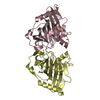
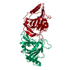
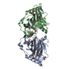
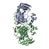
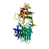
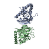
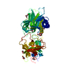
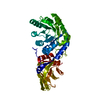
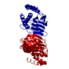
 Assembly
 Mass: 18.015 Da / Num. of mol.: 144 / Source method: isolated from a natural source / Formula: H2O
 Sample preparation
 / Beamline: X06DA / Wavelength: 1 Å
 Processing