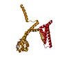
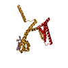
 Movie
Movie Controller


+ Open data

- Basic information
| Entry | Database: PDB / ID: 6yz2 | ||||||
|---|---|---|---|---|---|---|---|
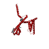
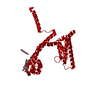
| Title | Full length Open-form Sodium Channel NavMs F208L | ||||||
 Components Components | Ion transport protein | ||||||
 Keywords Keywords | METAL TRANSPORT / Voltage Gated Sodium Channel / Membrane protein. | ||||||
| Function / homology |  Function and homology information Function and homology informationvoltage-gated sodium channel complex / voltage-gated sodium channel activity Similarity search - Function | ||||||
| Biological species |  Magnetococcus marinus (bacteria) Magnetococcus marinus (bacteria) | ||||||
| Method |  X-RAY DIFFRACTION / SYNCHROTRON / MOLECULAR REPLACEMENT / Resolution: 2.2 Å X-RAY DIFFRACTION / SYNCHROTRON / MOLECULAR REPLACEMENT / Resolution: 2.2 Å | ||||||
 Authors Authors | Sula, A. / Sait, L.G. / Hollingworth, D. / Wallace, B.A. | ||||||
| Funding support |  United Kingdom, 1items United Kingdom, 1items
| ||||||
 Citation Citation | Journal: Elife / Year: 2020 Title: Cannabidiol interactions with voltage-gated sodium channels. Authors: Sait, L.G. / Sula, A. / Ghovanloo, M.R. / Hollingworth, D. / Ruben, P.C. / Wallace, B.A. | ||||||
| History |
|
- Structure visualization
| Structure viewer | Molecule: MolmilJmol/JSmol |
|---|
- Downloads & links
-Download
| PDBx/mmCIF format | 6yz2.cif.gz | 121.6 KB | Display | PDBx/mmCIF format |
|---|---|---|---|---|
| PDB format | pdb6yz2.ent.gz | 93.5 KB | Display | PDB format |
| PDBx/mmJSON format | 6yz2.json.gz | Tree view | PDBx/mmJSON format | |
| Others |  Other downloads Other downloads |
-Validation report
| Arichive directory | https://data.pdbj.org/pub/pdb/validation_reports/yz/6yz2ftp://data.pdbj.org/pub/pdb/validation_reports/yz/6yz2 | HTTPS FTP |
|---|
-Related structure data
| Related structure data |  6yz0C 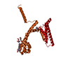 5hvxS S: Starting model for refinement C: citing same article ( |
|---|---|
| Similar structure data |


-Links
 PDBj
PDBj
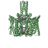
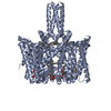
- Assembly
| Deposited unit | 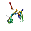
| ||||||||||||
|---|---|---|---|---|---|---|---|---|---|---|---|---|---|
| 1 | 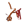
| ||||||||||||
| Unit cell |
| ||||||||||||
| Components on special symmetry positions |
|
-Components
| #1: Protein | Mass: 31128.680 Da / Num. of mol.: 1 / Mutation: F210L Source method: isolated from a genetically manipulated source Source: (gene. exp.) Magnetococcus marinus (strain ATCC BAA-1437 / JCM 17883 / MC-1) (bacteria)Gene: Mmc1_0798 / Production host: | ||||||||
|---|---|---|---|---|---|---|---|---|---|
| #2: Chemical |   Mass: 22.990 Da / Num. of mol.: 3 / Source method: obtained synthetically / Formula: Na Mass: 22.990 Da / Num. of mol.: 3 / Source method: obtained synthetically / Formula: Na#3: Chemical | 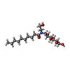  Mass: 379.489 Da / Num. of mol.: 2 / Source method: obtained synthetically / Formula: C18H37NO7 / Comment: detergent*YM Mass: 379.489 Da / Num. of mol.: 2 / Source method: obtained synthetically / Formula: C18H37NO7 / Comment: detergent*YM#4: Chemical |   Mass: 546.646 Da / Num. of mol.: 3 / Source method: obtained synthetically / Formula: C24H50O13 / Comment: precipitant*YM Mass: 546.646 Da / Num. of mol.: 3 / Source method: obtained synthetically / Formula: C24H50O13 / Comment: precipitant*YM#5: Water | ChemComp-HOH / |  Mass: 18.015 Da / Num. of mol.: 93 / Source method: isolated from a natural source / Formula: H2O Mass: 18.015 Da / Num. of mol.: 93 / Source method: isolated from a natural source / Formula: H2OHas ligand of interest | N | |
-Experimental details
-Experiment
| Experiment | Method: X-RAY DIFFRACTION / Number of used crystals: 1 |
|---|
- Sample preparation
| Crystal grow | Temperature: 277 K / Method: vapor diffusion, sitting drop / pH: 7 Details: 0.1M lithium sulphate, 0.1M HEPES, pH 7, and 40% v/v PEG200. |
|---|
-Data collection
| Diffraction | Mean temperature: 80 K / Serial crystal experiment: N |
|---|---|
| Diffraction source | Source: SYNCHROTRON / Site: EMBL/DESY, HAMBURG  / Beamline: X13 / Wavelength: 0.9797 Å / Beamline: X13 / Wavelength: 0.9797 Å |
| Detector | Type: DECTRIS PILATUS 6M / Detector: PIXEL / Date: Jul 2, 2019 |
| Radiation | Protocol: SINGLE WAVELENGTH / Monochromatic (M) / Laue (L): M / Scattering type: x-ray |
| Radiation wavelength | Wavelength: 0.9797 Å / Relative weight: 1 |
| Reflection | Resolution: 2.2→104.68 Å / Num. obs: 32388 / % possible obs: 100 % / Redundancy: 21.2 % / CC1/2: 0.999 / Net I/σ(I): 17.4 |
| Reflection shell | Resolution: 2.2→2.27 Å / Mean I/σ(I) obs: 2.2 / Num. unique obs: 2771 / CC1/2: 0.953 |
- Processing
| Software |
| ||||||||||||||||||||||||||||||||||||||||||||||||||||||||||||||||||||||||||||||||||||||||||||||||||||||||||||
|---|---|---|---|---|---|---|---|---|---|---|---|---|---|---|---|---|---|---|---|---|---|---|---|---|---|---|---|---|---|---|---|---|---|---|---|---|---|---|---|---|---|---|---|---|---|---|---|---|---|---|---|---|---|---|---|---|---|---|---|---|---|---|---|---|---|---|---|---|---|---|---|---|---|---|---|---|---|---|---|---|---|---|---|---|---|---|---|---|---|---|---|---|---|---|---|---|---|---|---|---|---|---|---|---|---|---|---|---|---|
| Refinement | Method to determine structure: MOLECULAR REPLACEMENT Starting model: 5hvx Resolution: 2.2→104.68 Å / Cor.coef. Fo:Fc: 0.895 / Cor.coef. Fo:Fc free: 0.887 / SU R Cruickshank DPI: 0.163 / Cross valid method: THROUGHOUT / σ(F): 0 / SU R Blow DPI: 0.164 / SU Rfree Blow DPI: 0.152 / SU Rfree Cruickshank DPI: 0.152
| ||||||||||||||||||||||||||||||||||||||||||||||||||||||||||||||||||||||||||||||||||||||||||||||||||||||||||||
| Displacement parameters | Biso max: 179.42 Å2 / Biso mean: 76.37 Å2 / Biso min: 30 Å2
| ||||||||||||||||||||||||||||||||||||||||||||||||||||||||||||||||||||||||||||||||||||||||||||||||||||||||||||
| Refine analyze | Luzzati coordinate error obs: 0.33 Å | ||||||||||||||||||||||||||||||||||||||||||||||||||||||||||||||||||||||||||||||||||||||||||||||||||||||||||||
| Refinement step | Cycle: final / Resolution: 2.2→104.68 Å
| ||||||||||||||||||||||||||||||||||||||||||||||||||||||||||||||||||||||||||||||||||||||||||||||||||||||||||||
| Refine LS restraints |
| ||||||||||||||||||||||||||||||||||||||||||||||||||||||||||||||||||||||||||||||||||||||||||||||||||||||||||||
| LS refinement shell | Resolution: 2.2→2.21 Å / Rfactor Rfree error: 0 / Total num. of bins used: 51
| ||||||||||||||||||||||||||||||||||||||||||||||||||||||||||||||||||||||||||||||||||||||||||||||||||||||||||||
| Refinement TLS params. | Method: refined / Origin x: 47.5502 Å / Origin y: 71.8823 Å / Origin z: 31.5119 Å
| ||||||||||||||||||||||||||||||||||||||||||||||||||||||||||||||||||||||||||||||||||||||||||||||||||||||||||||
| Refinement TLS group | Selection details: { A|* } |
